Research
Our lab develops computational imaging methods, which jointly design optics, devices, signal processing and algorithms, and enable novel capabilities that each one alone cannot. Our research is inherently interdisciplinary, combining expertise in optical engineering, physics and computation. We work on imaging technologies for biomedical, neuroscience, and semiconductor-related applications.
Computational Microscopy
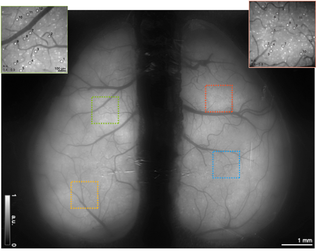 Computational Miniature Mesoscope (CM2)
We develop “wearable” computational fluorescence microscope that achieves cm-scale FOV and µm-scale resolution with single-shot 3D imaging capability. A complete publication list is here.
-
-
-
-
-
-
-
-
- Xue, et al, Single-Shot 3D Widefield Fluorescence Imaging with a Computational Miniature Mesoscope, Science Advances 21 2020: EABB7508
- Xue, et al, Deep-learning-augmented computational miniature mesoscope, Optica 9, 1009-1021 (2022).
- Yang, et al, Wide-field, high-resolution reconstruction in computational multi-aperture miniscope using a Fourier neural network, Optica 11, 860-871 (2024).
-
-
-
-
-
-
-
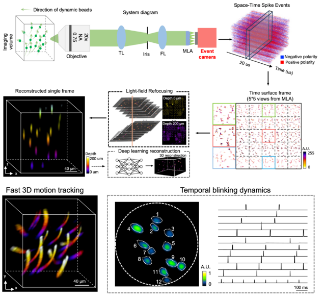
Event-driven dynamic microscopy
We develop computational “event-driven” microscopy techniques to address challenges in dynamic imaging.
-
-
-
-
-
-
-
-
- Guo, Ruipeng, et al. Dual-channel event microscopy for ultrafast biological imaging, PNAS 2026.
- Guo, Ruipeng, et al. EventLFM: Event Camera integrated Fourier Light Field Microscopy for Ultrafast 3D imaging. Light: Science & Applications 2024.
-
-
-
-
-
-
-
Computational Metrology
We developed computational imaging techniques for semiconductor metrology and inspection applications. A complete publication list is here.
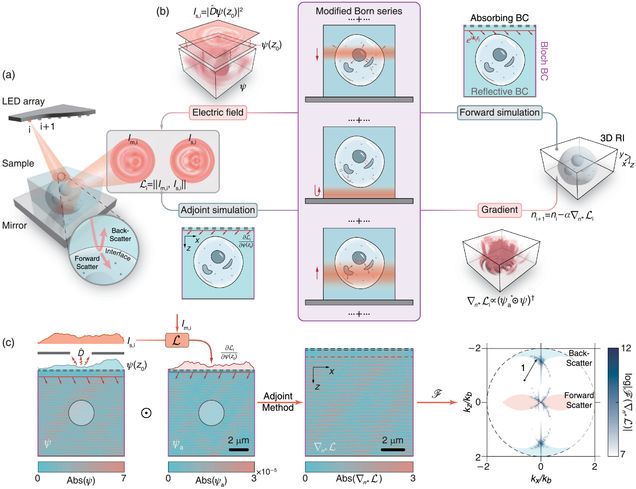 Substrate-Enhanced Diffraction Tomography
We develop a reflection-mode diffraction tomography technique that enables the simultaneous recovery of forward- and backward-scattering information for high-resolution 3D refractive index reconstruction.
-
-
-
-
-
-
-
-
- T. Li, et. al. Transfer-function approach to substrate-enhanced diffraction tomography, Optica, 2026.
- J. Zhu, et. al. Reflection-mode Multi-slice Fourier Ptychographic Tomography, IEEE Transactions on Computational Imaging, 2026.
- T. Li, et. al. Reflection-mode diffraction tomography of multiple-scattering samples on a reflective substrate from intensity images, Optica, 2025.
-
-
-
-
-
-
-
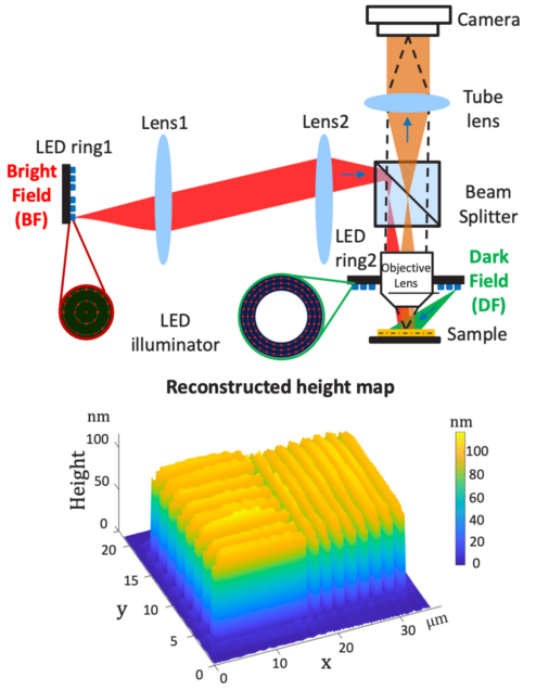
Fourier Ptychograhpic Topography
We develop novel topography techniques based on the reflection-mode Fourier ptychographic microscopy, termed Fourier ptychograhpic topography (FPT). FPT provides both a wide FOV and high resolution, and achieves nanoscale height reconstruction accuracy.
-
-
-
-
-
-
-
-
- Y. Shen, et. al. Dual-wavelength Fourier Ptychographic Topography, IEEE Transactions on Computational Imaging, 2026.
- H. Wang, et. al. Fourier ptychographic topography, Optics Express 31, 11007-11018 (2023).
-
-
-
-
-
-
-
Deep Learning for Computational Imaging
 Diffusion models
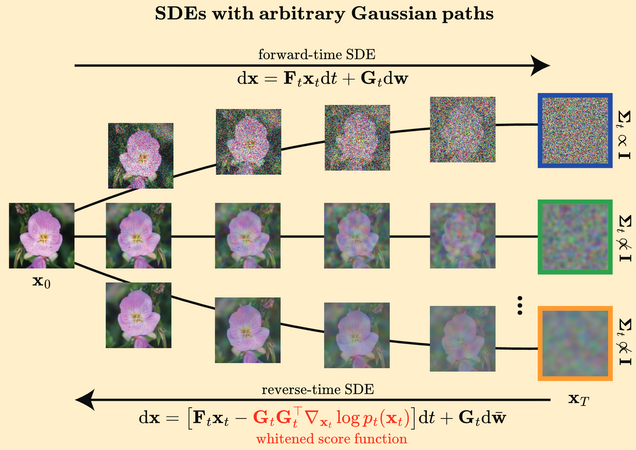
We work on diffusion models for solving inverse problems.
-
-
-
-
-
-
-
-
- Alido, et al, Whitened Score Diffusion: A Structured Prior for Imaging Inverse Problems, NeurIPS, 2025.
-
-
-
-
-
-
-
Neural fields
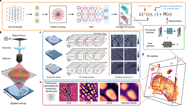
We develop computational imaging techniques that leverage implicit neural representations (neural fields).
-
-
-
-
-
-
-
-
- Wang, et al, NeuPh: scalable and generalizable neural phase retrieval with local conditional neural fields, Advanced Photonics Nexus 3, 056005 (2024).
- Liu, et al, Recovery of Continuous 3D Refractive Index Maps from Discrete Intensity-Only Measurements using Neural Fields, Nature Machine Intelligence 4, 781–791 (2022).
-
-
-
-
-
-
-
Reliable deep learning with Uncertainty Quantification
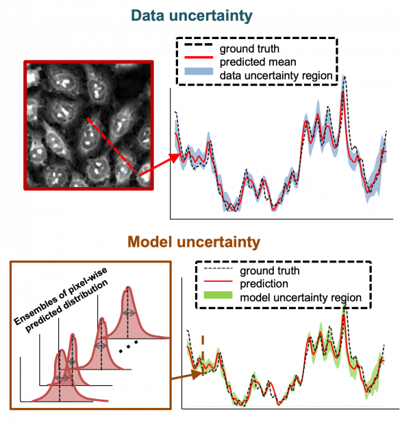
We develop uncertainty quantification techniques to provide more reliable deep learning predictions for quantitative bio-imaging.
-
-
-
-
-
-
-
-
- Xue, et al, Reliable deep learning-based phase imaging with uncertainty quantification, Optica 6, 618-629 (2019).
- Liu,, et al. Deep spectral learning for label-free optical imaging oximetry with uncertainty quantification. Light: Science & Applications 8.1 (2019): 102.
-
-
-
-
-
-
-
Deep learning for Quantitative Bio-imaging
We work in the following major directions. A complete publication list is here.
 Physics-informed deep learning
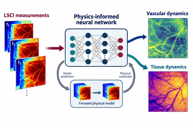
We develop physics-informed deep learning techniques that integrate physical models with neural networks for fast and interpretable quantitative biomedical imaging.
-
-
-
-
-
-
-
-
- Li, et al., Physics-informed neural network for mapping vascular and tissue dynamics using laser speckle contrast imaging, Neurophotonics 13, 035012 (2026).
-
-
-
-
-
-
-
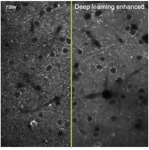
Deep learning for in-vivo neural signal modeling & extraction
We develop various deep learning techniques to enhance and analyze in-vivo neural signals. A complete publication list is here.
-
-
-
-
-
-
-
-
- Chang, et al, DeepVID v2: self-supervised denoising with decoupled spatiotemporal enhancement for low-photon voltage imaging, Neurophotonics, 2024.
- Platisa, et al, High-speed low-light in vivo two-photon voltage imaging of large neuronal populations, Nature Methods 20, pages1095–1103 (2023)
-
-
-
-
-
-
-
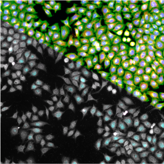
Cross-modality information transfer
We work on deep learning technique that allows knowledge transfer across different bio-imaging modalities.
-
-
-
-
-
-
-
-
- Cheng, et al, Single-cell cytometry via multiplexed fluorescence prediction by label-free reflectance microscopy, Science Advances 15 Jan 2021: Vol. 7, no. 3, eabe0431.
-
-
-
-
-
-
-
Computational Phase Imaging
We work in the following major directions.
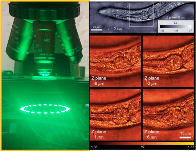
Intensity Diffraction Tomography
We are developing high-speed computational 3D phase microscopy techniques by leveraging simple optical setups and advanced algorithms.
-
-
-
-
-
-
-
-
- Li, et al, High-speed in vitro intensity diffraction tomography, Advanced Photonics, 1(6), 066004 (2019).
-
-
-
-
-
-
-
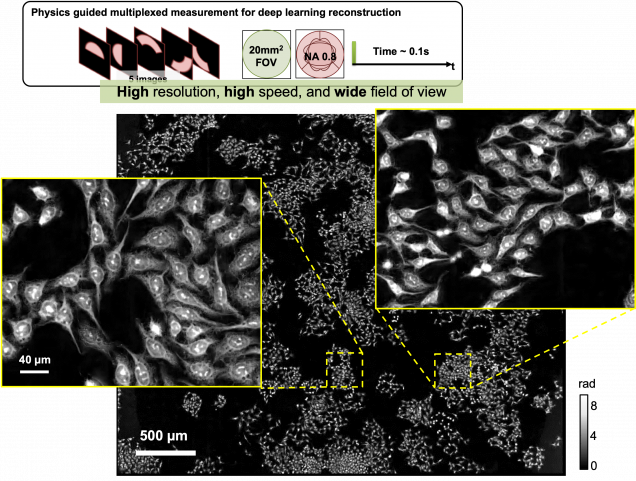
Fourier Ptychography
We are working computational microscopy techniques to achieve gigapixel phase imaging.
-
-
-
-
-
-
-
-
- Tian, et al, Computational illumination for high-speed in vitro Fourier ptychographic microscopy, Optica 2(10), 904-911 (2015).
-
-
-
-
-
-
-