Computational Phase Microscopy

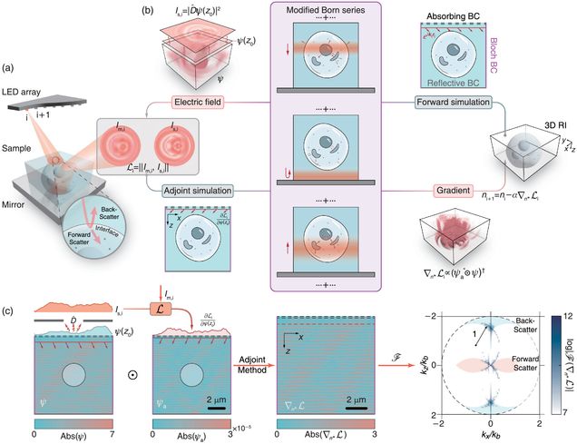
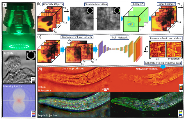
Multiple-scattering simulator-trained neural network for intensity diffraction tomography
A. Matlock, J. Zhu, L. Tian
Optics Express 31, 4094-4107 (2023)
Bond-Selective Intensity Diffraction Tomography
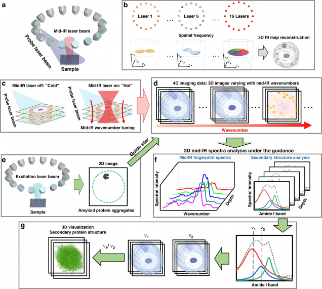
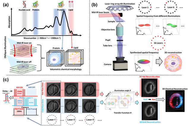
Jian Zhao, Alex Matlock, Hongbo Zhu, Ziqi Song, Jiabei Zhu, Biao Wang, Fukai Chen, Yuewei Zhan, Zhicong Chen, Yihong Xu, Xingchen Lin, Lei Tian, Ji-Xin Cheng
Nat Commun 13, 7767 (2022).

Recovery of Continuous 3D Refractive Index Maps from Discrete Intensity-Only Measurements using Neural Fields
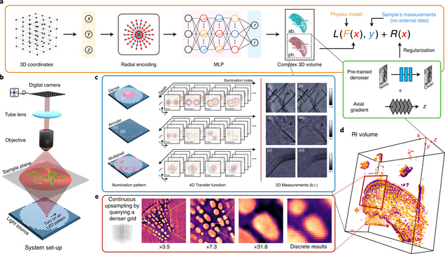
Renhao Liu, Yu Sun, Jiabei Zhu, Lei Tian, Ulugbek Kamilov
Nature Machine Intelligence 4, 781–791 (2022).
High-fidelity intensity diffraction tomography with a non-paraxial multiple-scattering model
Jiabei Zhu, Hao Wang, Lei Tian
Optics Express Vol. 30, Issue 18, pp. 32808-32821 (2022).
 Github Project


SIMBA: Scalable Inversion in Optical Tomography using Deep Denoising Priors
Zihui Wu, Yu Sun, Alex Matlock, Jiaming Liu, Lei Tian, Ulugbek S. Kamilov
IEEE Journal of Selected Topics in Signal Processing 14(6), 2020.

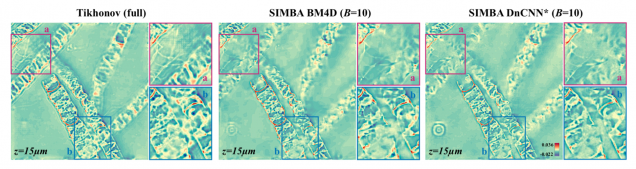
Two features desired in a three-dimensional (3D) optical tomographic image reconstruction algorithm are the ability to reduce imaging artifacts and to do fast processing of large data volumes. Traditional iterative inversion algorithms are impractical in this context due to their heavy computational and memory requirements. We propose and experimentally validate a novel scalable iterative minibatch algorithm (SIMBA) for fast and high-quality optical tomographic imaging. SIMBA enables high-quality imaging by combining two complementary information sources: the physics of the imaging system characterized by its forward model and the imaging prior characterized by a denoising deep neural net. SIMBA easily scales to very large 3D tomographic datasets by processing only a small subset of measurements at each iteration. We establish the theoretical fixed-point convergence of SIMBA under nonexpansive denoisers for convex data-fidelity terms. We validate SIMBA on both simulated and experimentally collected intensity diffraction tomography (IDT) datasets. Our results show that SIMBA can significantly reduce the computational burden of 3D image formation without sacrificing the imaging quality.

High-Throughput, High-Resolution Interferometric Light Microscopy of Biological Nanoparticles
C. Yurdakul, O. Avci, A. Matlock, A. J Devaux, M. V Quintero, E. Ozbay, R. A Davey, J. H Connor, W C. Karl, L. Tian, M S. Ünlü
ACS Nano 2020, 14, 2, 2002-2013

Label-free, visible light microscopy is an indispensable tool for studying biological nanoparticles (BNPs). However, conventional imaging techniques have two major challenges: (i) weak contrast due to low-refractive-index difference with the surrounding medium and exceptionally small size and (ii) limited spatial resolution. Advances in interferometric microscopy have overcome the weak contrast limitation and enabled direct detection of BNPs, yet lateral resolution remains as a challenge in studying BNP morphology. Here, we introduce a wide-field interferometric microscopy technique augmented by computational imaging to demonstrate a 2-fold lateral resolution improvement over a large field-of-view (>100 × 100 μm2), enabling simultaneous imaging of more than 104 BNPs at a resolution of ∼150 nm without any labels or sample preparation. We present a rigorous vectorial-optics-based forward model establishing the relationship between the intensity images captured under partially coherent asymmetric illumination and the complex permittivity distribution of nanoparticles. We demonstrate high-throughput morphological visualization of a diverse population of Ebola virus-like particles and a structurally distinct Ebola vaccine candidate. Our approach offers a low-cost and robust label-free imaging platform for high-throughput and high-resolution characterization of a broad size range of BNPs.

LED array reflectance microscopy for scattering-based multi-contrast imaging
Weiye Song, Alex Matlock, Sipei Fu, Xiaodan Qin, Hui Feng, Christopher V. Gabel, Lei Tian, and Ji Yi
Opt. Lett. 45, 1647-1650 (2020)

LED array microscopy is an emerging platform for computational imaging with significant utility for biological imaging. Existing LED array systems often exploit transmission imaging geometries of standard brightfield microscopes that leave the rich backscattered field undetected. This backscattered signal contains high-resolution sample information with superb sensitivity to subtle structural features that make it ideal for biological sensing and detection. Here, we develop an LED array reflectance microscope capturing the sample’s backscattered signal. In particular, we demonstrate multimodal brightfield, darkfield, and differential phase contrast imaging on fixed and living biological specimens including Caenorhabditis elegans (C. elegans), zebrafish embryos, and live cell cultures. Video-rate multimodal imaging at 20 Hz records real time features of freely moving C. elegans and the fast beating heart of zebrafish embryos. Our new reflectance mode is a valuable addition to the LED array microscopic toolbox.

Inverse scattering for reflection intensity phase microscopy
Alex Matlock, Anne Sentenac, Patrick C. Chaumet, Ji Yi, and Lei Tian
Biomedical Optics Express 11, pp. 911-926 (2020).

 Github Project

Reflection phase imaging provides label-free, high-resolution characterization of biological samples, typically using interferometric-based techniques. Here, we investigate reflection phase microscopy from intensity-only measurements under diverse illumination. We evaluate the forward and inverse scattering model based on the first Born approximation for imaging scattering objects above a glass slide. Under this design, the measured field combines linear forward-scattering and height-dependent nonlinear back-scattering from the object that complicates object phase recovery. Using only the forward-scattering, we derive a linear inverse scattering model and evaluate this model’s validity range in simulation and experiment using a standard reflection microscope modified with a programmable light source. Our method provides enhanced contrast of thin, weakly scattering samples that complement transmission techniques. This model provides a promising development for creating simplified intensity-based reflection quantitative phase imaging systems easily adoptable for biological research.

High-speed in vitro intensity diffraction tomography
Jiaji Li, Alex Matlock, Yunzhe Li, Qian Chen, Chao Zuo, Lei Tian
Advanced Photonics, 1(6), 066004 (2019).
on the cover story
⭑ Highlighted at Programmable LED ring enables label-free 3D tomography for conventional microscopes

 Github Project

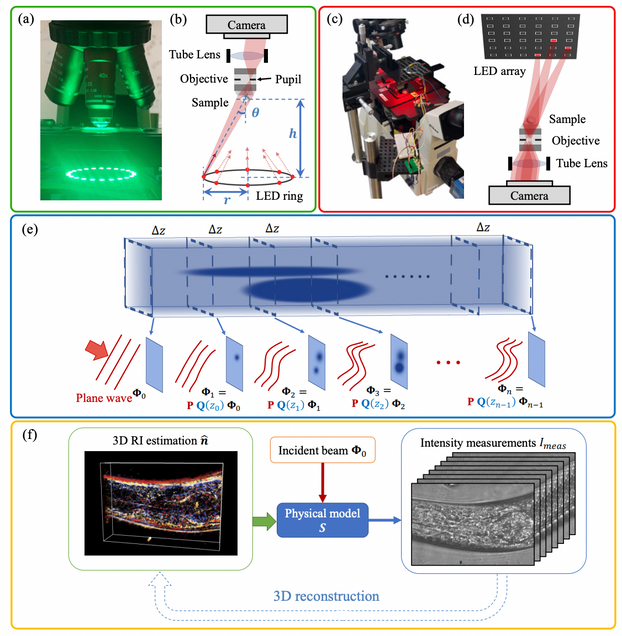
We demonstrate a label-free, scan-free intensity diffraction tomography technique utilizing annular illumination (aIDT) to rapidly characterize large-volume 3D refractive index distributions in vitro. By optimally matching the illumination geometry to the microscope pupil, our technique reduces the data requirement by 60× to achieve high-speed 10 Hz volume rates. Using 8 intensity images, we recover 350×100×20μm3 volumes with near diffraction-limited lateral resolution of 487 nm and axial resolution of 3.4 μm. Our technique’s large volume rate and high resolution enables 3D quantitative phase imaging of complex living biological samples across multiple length scales. We demonstrate aIDT’s capabilities on unicellular diatom microalgae, epithelial buccal cell clusters with native bacteria, and live Caenorhabditis elegans specimens. Within these samples, we recover macroscale cellular structures, subcellular organelles, and dynamic micro-organism tissues with minimal motion artifacts. Quantifying such features has significant utility in oncology, immunology, and cellular pathophysiology, where these morphological features are evaluated for changes in the presence of disease, parasites, and new drug treatments. aIDT shows promise as a powerful high-speed, label-free microscopy technique for these applications where natural imaging is required to evaluate environmental effects on a sample in real-time.

High-throughput, volumetric quantitative phase imaging with multiplexed intensity diffraction tomography
Alex Matlock, Lei Tian
Biomed. Opt. Express 10, pp. 6432-6448 (2019).

Intensity diffraction tomography (IDT) provides quantitative, volumetric refractive index reconstructions of unlabeled biological samples from intensity-only measurements. IDT is scanless and easily implemented in standard optical microscopes using an LED array but suffers from large data requirements and slow acquisition speeds. Here, we develop multiplexed IDT (mIDT), a coded illumination framework providing high volume-rate IDT for evaluating dynamic biological samples. mIDT combines illuminations from an LED grid using physical model-based design choices to improve acquisition rates and reduce dataset size with minimal loss to resolution and reconstruction quality. We analyze the optimal design scheme with our mIDT framework in simulation using the reconstruction error compared to conventional IDT and theoretical acquisition speed. With the optimally determined mIDT scheme, we achieve hardware-limited 4Hz acquisition rates enabling 3D refractive index distribution recovery on live Caenorhabditis elegans worms and embryos as well as epithelial buccal cells. Our mIDT architecture provides a 60 × speed improvement over conventional IDT and is robust across different illumination hardware designs, making it an easily adoptable imaging tool for volumetrically quantifying biological samples in their natural state.

Reliable deep learning-based phase imaging with uncertainty quantification
Yujia Xue, Shiyi Cheng, Yunzhe Li, Lei Tian
Optica 6, 618-629 (2019).

 Github Project

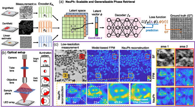
Emerging deep-learning (DL)-based techniques have significant potential to revolutionize biomedical imaging. However, one outstanding challenge is the lack of reliability assessment in the DL predictions, whose errors are commonly revealed only in hindsight. Here, we propose a new Bayesian convolutional neural network (BNN)-based framework that overcomes this issue by quantifying the uncertainty of DL predictions. Foremost, we show that BNN-predicted uncertainty maps provide surrogate estimates of the true error from the network model and measurement itself. The uncertainty maps characterize imperfections often unknown in real-world applications, such as noise, model error, incomplete training data, and out-of-distribution testing data. Quantifying this uncertainty provides a per-pixel estimate of the confidence level of the DL prediction as well as the quality of the model and data set. We demonstrate this framework in the application of large space–bandwidth product phase imaging using a physics-guided coded illumination scheme. From only five multiplexed illumination measurements, our BNN predicts gigapixel phase images in both static and dynamic biological samples with quantitative credibility assessment. Furthermore, we show that low-certainty regions can identify spatially and temporally rare biological phenomena. We believe our uncertainty learning framework is widely applicable to many DL-based biomedical imaging techniques for assessing the reliability of DL predictions.

Deep learning approach to Fourier ptychographic microscopy
Thanh Nguyen, Yujia Xue, Yunzhe Li, Lei Tian, George Nehmetallah
Opt. Express 26, 26470-26484 (2018).

Convolutional neural networks (CNNs) have gained tremendous success in solving complex inverse problems. The aim of this work is to develop a novel CNN framework to reconstruct video sequence of dynamic live cells captured using a computational microscopy technique, Fourier ptychographic microscopy (FPM). The unique feature of the FPM is its capability to reconstruct images with both wide field-of-view (FOV) and high resolution, i.e. a large space-bandwidth-product (SBP), by taking a series of low resolution intensity images. For live cell imaging, a single FPM frame contains thousands of cell samples with different morphological features. Our idea is to fully exploit the statistical information provided by this large spatial ensemble so as to make predictions in a sequential measurement, without using any additional temporal dataset. Specifically, we show that it is possible to reconstruct high-SBP dynamic cell videos by a CNN trained only on the first FPM dataset captured at the beginning of a time-series experiment. Our CNN approach reconstructs a 12800X10800 pixels phase image using only ~25 seconds, a 50X speedup compared to the model-based FPM algorithm. In addition, the CNN further reduces the required number of images in each time frame by ~6X. Overall, this significantly improves the imaging throughput by reducing both the acquisition and computational times. The proposed CNN is based on the conditional generative adversarial network (cGAN) framework. Additionally, we also exploit transfer learning so that our pre-trained CNN can be further optimized to image other cell types. Our technique demonstrates a promising deep learning approach to continuously monitor large live-cell populations over an extended time and gather useful spatial and temporal information with sub-cellular resolution.

patternAndROI

High-throughput intensity diffraction tomography with a computational microscope
Ruilong Ling, Waleed Tahir, Hsing-Ying Lin, Hakho Lee, and Lei Tian
Biomed. Opt. Express 9, 2130-2141 (2018).

 Github Project

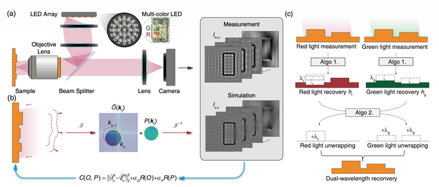
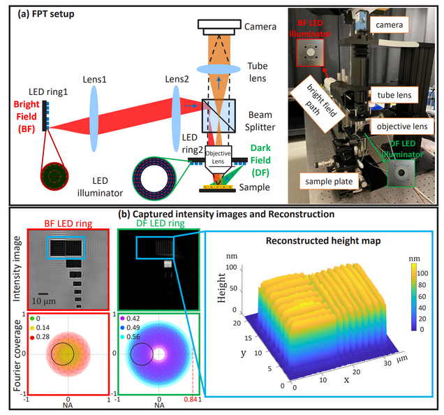
We demonstrate a motion-free intensity diffraction tomography technique that enables the direct inversion of 3D phase and absorption from intensity-only measurements for weakly scattering samples. We derive a novel linear forward model featuring slice-wise phase and absorption transfer functions using angled illumination. This new framework facilitates flexible and efficient data acquisition, enabling arbitrary sampling of the illumination angles. The reconstruction algorithm performs 3D synthetic aperture using a robust computation and memory efficient slice-wise deconvolution to achieve resolution up to the incoherent limit. We demonstrate our technique with thick biological samples having both sparse 3D structures and dense cell clusters. We further investigate the limitation of our technique when imaging strongly scattering samples. Imaging performance and the influence of multiple scattering is evaluated using a 3D sample consisting of stacked phase and absorption resolution targets. This computational microscopy system is directly built on a standard commercial microscope with a simple LED array source add-on, and promises broad applications by leveraging the ubiquitous microscopy platforms with minimal hardware modifications.

IDT


3D differential phase contrast microscopy

Michael Chen, Lei Tian, Laura Waller
Biomed. Opt. Express 7, 3940-3950 (2016).

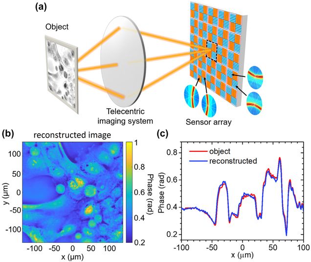
We demonstrate 3D phase and absorption recovery from partially coherent intensity images captured with a programmable LED array source. Images are captured through-focus with four different illumination patterns. Using first Born and weak object approximations (WOA), a linear 3D differential phase contrast (DPC) model is derived. The partially coherent transfer functions relate the sample’s complex refractive index distribution to intensity measurements at varying defocus. Volumetric reconstruction is achieved by a global FFT-based method, without an intermediate 2D phase retrieval step. Because the illumination is spatially partially coherent, the transverse resolution of the reconstructed field achieves twice the NA of coherent systems and improved axial resolution.

3D qDPC

Computational illumination for high-speed in vitro Fourier ptychographic microscopy
L. Tian, Z. Liu, L. Yeh, M. Chen, J. Zhong, L. Waller
Optica 2(10), 904-911 (2015).

We demonstrate a new computational illumination technique that achieves a large space-bandwidth-time product, for quantitative phase imaging of unstained live samples in vitro. Microscope lenses can have either a large field of view (FOV) or high resolution, and not both. Fourier ptychographic microscopy (FPM) is a new computational imaging technique that circumvents this limit by fusing information from multiple images taken with different illumination angles. The result is a gigapixel-scale image having both a wide FOV and high resolution, i.e., a large space-bandwidth product. FPM has enormous potential for revolutionizing microscopy and has already found application in digital pathology. However, it suffers from long acquisition times (of the order of minutes), limiting throughput. Faster capture times would not only improve the imaging speed, but also allow studies of live samples, where motion artifacts degrade results. In contrast to fixed (e.g., pathology) slides, live samples are continuously evolving at various spatial and temporal scales. Here, we present a new source coding scheme, along with real-time hardware control, to achieve 0.8 NA resolution across a 4x FOV with subsecond capture times. We propose an improved algorithm and a new initialization scheme, which allow robust phase reconstruction over long time-lapse experiments. We present the first FPM results for both growing and confluent in vitro cell cultures, capturing videos of subcellular dynamical phenomena in popular cell lines undergoing division and migration. Our method opens up FPM to applications with live samples, for observing rare events in both space and time.

InvitroFPM

3D intensity and phase imaging from light field measurements in an LED array microscope
Lei Tian, L. Waller
Optica 2, 104-111 (2015).
 the 15 Most Cited Articles in Optica published in 2015 (Source: OSA, 2019)

Realizing high resolution across large volumes is challenging for 3D imaging techniques with high-speed acquisition. Here, we describe a new method for 3D intensity and phase recovery from 4D light field measurements, achieving enhanced resolution via Fourier Ptychography. Starting from geometric optics light field refocusing, we incorporate phase retrieval and correct diffraction artifacts. Further, we incorporate dark-field images to achieve lateral resolution beyond the diffraction limit of the objective (5x larger NA) and axial resolution better than the depth of field, using a low magnification objective with a large field of view. Our iterative reconstruction algorithm uses a multi-slice coherent model to estimate the 3D complex transmittance function of the sample at multiple depths, without any weak or single-scattering approximations. Data is captured by an LED array microscope with computational illumination, which enables rapid scanning of angles for fast acquisition. We demonstrate the method with thick biological samples in a modified commercial microscope, indicating the technique’s versatility for a wide range of applications.

3DFourierPtychography

Multiplexed coded illumination for Fourier Ptychography with an LED array microscope
Lei Tian, X. Li, K. Ramchandran, L. Waller
Biomedical Optics Express 5, 2376-2389 (2014).
 the decade’s most highly cited Articles in Biomed. Opt. Express (Source: OSA, 2020)
⭑ Highly cited (Top 1%) papers between 2008-2018 (source: Web of Science, 2019)

Fourier Ptychography is a new computational microscopy technique that achieves gigapixel images with both wide field of view and high resolution in both phase and amplitude. The hardware setup involves a simple replacement of the microscope’s illumination unit with a programmable LED array, allowing one to flexibly pattern illumination angles without any moving parts. In previous work, a series of low-resolution images was taken by sequentially turning on each single LED in the array, and the data were then combined to recover a bandwidth much higher than the one allowed by the original imaging system. Here, we demonstrate a multiplexed illumination strategy in which multiple randomly selected LEDs are turned on for each image. Since each LED corresponds to a different area of Fourier space, the total number of images can be significantly reduced, without sacrificing image quality. We demonstrate this method experimentally in a modified commercial microscope. Compared to sequential scanning, our multiplexed strategy achieves similar results with approximately an order of magnitude reduction in both acquisition time and data capture requirements.

MultiplexFP

Quantitative differential phase contrast imaging in an LED array microscope
L. Tian, L. Waller
Opt. Express 23, 11394-11403 (2015).

Illumination-based differential phase contrast (DPC) is a phase imaging method that uses a pair of images with asymmetric illumination patterns. Distinct from coherent techniques, DPC relies on spatially partially coherent light, providing 2× better lateral resolution, better optical sectioning and immunity to speckle noise. In this paper, we derive the 2D weak object transfer function (WOTF) and develop a quantitative phase reconstruction method that is robust to noise. The effect of spatial coherence is studied experimentally, and multiple-angle DPC is shown to provide improved frequency coverage for more stable phase recovery. Our method uses an LED array microscope to achieve real-time (10 Hz) quantitative phase imaging with in vitro live cell samples.

DPC

3D differential phase contrast microscopy with computational illumination using an LED array
Lei Tian, J. Wang, L. Waller
Optics Letters 39, 1326 – 1329 (2014).

We demonstrate 3D differential phase-contrast (DPC) microscopy, based on computational illumination with a programmable LED array. By capturing intensity images with various illumination angles generated by sequentially patterning an LED array source, we digitally refocus images through various depths via light field processing. The intensity differences from images taken at complementary illumination angles are then used to generate DPC images, which are related to the gradient of phase. The proposed method achieves 3D DPC with simple, inexpensive optics and no moving parts. We experimentally demonstrate our method by imaging a camel hair sample in 3D.

3DDPC


Experimental robustness of Fourier Ptychography phase retrieval algorithms
L. Yeh, J. Dong, J. Zhong, L. Tian, M. Chen, G. Tang, M. Soltanolkotabi, L. Waller
Opt. Express 23(26) 33212-33238 (2015).

Fourier ptychography is a new computational microscopy technique that provides gigapixel-scale intensity and phase images with both wide field-of-view and high resolution. By capturing a stack of low-resolution images under different illumination angles, an inverse algorithm can be used to computationally reconstruct the high-resolution complex field. Here, we compare and classify multiple proposed inverse algorithms in terms of experimental robustness. We find that the main sources of error are noise, aberrations and mis-calibration (i.e. model mis-match). Using simulations and experiments, we demonstrate that the choice of cost function plays a critical role, with amplitude-based cost functions performing better than intensity-based ones. The reason for this is that Fourier ptychography datasets consist of images from both brightfield and darkfield illumination, representing a large range of measured intensities. Both noise (e.g. Poisson noise) and model mis-match errors are shown to scale with intensity. Hence, algorithms that use an appropriate cost function will be more tolerant to both noise and model mis-match. Given these insights, we propose a global Newton’s method algorithm which is robust and accurate. Finally, we discuss the impact of procedures for algorithmic correction of aberrations and mis-calibration.

FPM_algorithm

Adaptive 3D descattering with a dynamic synthesis network
Waleed Tahir, Hao Wang, Lei Tian
Light: Science & Applications 11, 42, 2022

 Github Project

Deep learning has been broadly applied to imaging in scattering applications. A common framework is to train a “descattering” neural network for image recovery by removing scattering artifacts. To achieve the best results on a broad spectrum of scattering conditions, individual “expert” networks have to be trained for each condition. However, the performance of the expert sharply degrades when the scattering level at the testing time differs from the training. An alternative approach is to train a “generalist” network using data from a variety of scattering conditions. However, the generalist generally suffers from worse performance as compared to the expert trained for each scattering condition. Here, we develop a drastically different approach, termed dynamic synthesis network (DSN), that can dynamically adjust the model weights and adapt to different scattering conditions. The adaptability is achieved by a novel architecture that enables dynamically synthesizing a network by blending multiple experts using a gating network. Notably, our DSN adaptively removes scattering artifacts across a continuum of scattering conditions regardless of whether the condition has been used for the training, and consistently outperforms the generalist. By training the DSN entirely on a multiple-scattering simulator, we experimentally demonstrate the network’s adaptability and robustness for 3D descattering in holographic 3D particle imaging. We expect the same concept can be adapted to many other imaging applications, such as denoising, and imaging through scattering media. Broadly, our dynamic synthesis framework opens up a new paradigm for designing highly adaptive deep learning and computational imaging techniques.

Roadmap on digital holography
Bahram Javidi, Artur Carnicer, Arun Anand, George Barbastathis, Wen Chen, Pietro Ferraro, J. W. Goodman, Ryoichi Horisaki, Kedar Khare, Malgorzata Kujawinska, Rainer A. Leitgeb, Pierre Marquet, Takanori Nomura, Aydogan Ozcan, YongKeun Park, Giancarlo Pedrini, Pascal Picart, Joseph Rosen, Genaro Saavedra, Natan T. Shaked, Adrian Stern, Enrique Tajahuerce, Lei Tian, Gordon Wetzstein, and Masahiro Yamaguchi
Optics Express Vol. 29, Issue 22, pp. 35078-35118 (2021).

This Roadmap article on digital holography provides an overview of a vast array of research activities in the field of digital holography. The paper consists of a series of 25 sections from the prominent experts in digital holography presenting various aspects of the field on sensing, 3D imaging and displays, virtual and augmented reality, microscopy, cell identification, tomography, label-free live cell imaging, and other applications. Each section represents the vision of its author to describe the significant progress, potential impact, important developments, and challenging issues in the field of digital holography.

Large-scale holographic particle 3D imaging with the beam propagation model
Hao Wang, Waleed Tahir, Jiabei Zhu, Lei Tian
Opt. Express 29, 17159-17172 (2021)
 Github Project

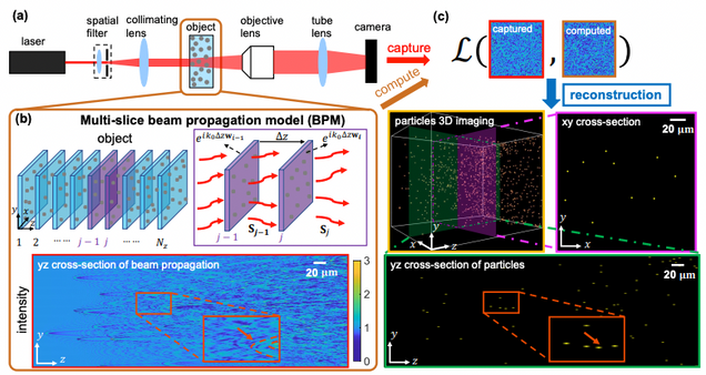
We develop a novel algorithm for large-scale holographic reconstruction of 3D particle fields. Our method is based on a multiple-scattering beam propagation method (BPM) combined with sparse regularization that enables recovering dense 3D particles of high refractive index contrast from a single hologram. We show that the BPM-computed hologram generates intensity statistics closely matching with the experimental measurements and provides up to 9× higher accuracy than the single-scattering model. To solve the inverse problem, we devise a computationally efficient algorithm, which reduces the computation time by two orders of magnitude as compared to the state-of-the-art multiple-scattering-based technique. We demonstrate superior reconstruction accuracy in both simulations and experiments under different scattering strengths. We show that the BPM reconstruction significantly outperforms the single-scattering method in particular for deep imaging depths and high particle densities.

Holographic particle-localization under multiple scattering
Waleed Tahir, Ulugbek S. Kamilov, Lei Tian
Advanced Photonics, 1(3), 036003 (2019).

We introduce a computational framework that incorporates multiple scattering for large-scale three-dimensional (3-D) particle localization using single-shot in-line holography. Traditional holographic techniques rely on single-scattering models that become inaccurate under high particle densities and large refractive index contrasts. Existing multiple scattering solvers become computationally prohibitive for large-scale problems, which comprise millions of voxels within the scattering volume. Our approach overcomes the computational bottleneck by slicewise computation of multiple scattering under an efficient recursive framework. In the forward model, each recursion estimates the next higher-order multiple scattered field among the object slices. In the inverse model, each order of scattering is recursively estimated by a nonlinear optimization procedure. This nonlinear inverse model is further supplemented by a sparsity promoting procedure that is particularly effective in localizing 3-D distributed particles. We show that our multiple-scattering model leads to significant improvement in the quality of 3-D localization compared to traditional methods based on single scattering approximation. Our experiments demonstrate robust inverse multiple scattering, allowing reconstruction of 100 million voxels from a single 1-megapixel hologram with a sparsity prior. The performance bound of our approach is quantified in simulation and validated experimentally. Our work promises utilization of multiple scattering for versatile large-scale applications.

Compressive holographic video
Zihao Wang, Leonidas Spinoulas, Kuan He, Lei Tian, Oliver Cossairt, Aggelos K. Katsaggelos, and Huaijin Chen
Opt. Express 25, 250-262 (2017).

Compressed sensing has been discussed separately in spatial and temporal domains. Compressive holography has been introduced as a method that allows 3D tomographic reconstruction at different depths from a single 2D image. Coded exposure is a temporal compressed sensing method for high speed video acquisition. In this work, we combine compressive holography and coded exposure techniques and extend the discussion to 4D reconstruction in space and time from one coded captured image. In our prototype, digital in-line holography was used for imaging macroscopic, fast moving objects. The pixel-wise temporal modulation was implemented by a digital micromirror device. In this paper we demonstrate 10× temporal super resolution with multiple depths recovery from a single image. Two examples are presented for the purpose of recording subtle vibrations and tracking small particles within 5 ms.

dhv

Compressive holographic two-dimensional localization with 1/302 subpixel accuracy
Y. Liu, Lei Tian, C. Hsieh, G. Barbastathis
Optics Express 22, 9774-9782 (2014).

We propose the use of compressive holography for two–dimensional (2D) subpixel motion localization. Our approach is based on computational implementation of edge–extraction using a Fourier–plane spiral phase mask, followed by compressive reconstruction of the edge of the object. Using this technique and relatively low–cost computer and piezo motion stage to establish ground truth for the motion, we demonstrated localization within 1/30th of a camera pixel in each linear dimension.

subpixel2D

Scanning-free compressive holography for object localization with subpixel accuracy
Y. Liu, Lei Tian, J. W. Lee, H. Y. H. Huang, M. S. Triantafyllou, G. Barbastathis
Optics Letters 37, 3357-3359 (2012).

We propose quantitative localization measurement of a known object with subpixel accuracy using compressive holography. We analyze the theoretical optimal solution in the compressive sampling framework and experimentally demonstrate localization accuracy of 1/45 pixel, in good agreement with the analysis.

subpixel1D

Quantitative measurement of size and three-dimensional position of fast moving bubbles in air-water mixture flows using digital holography
Lei Tian, N. Loomis, J. Dominguez-Caballero, G. Barbastathis
Applied Optics 49, 1549 (2010).

We present a digital in-line holographic imaging system for measuring the size and three-dimensional position of fast-moving bubbles in air–water mixture flows. The captured holograms are numerically processed by performing a two-dimensional projection followed by local depth estimation to quickly and efficiently obtain the size and position information of multiple bubbles simultaneously. Statistical analysis on measured bubble size distributions shows that they follow lognormal or gamma distributions.

BUBBLE