Optical neural signal modeling & extraction
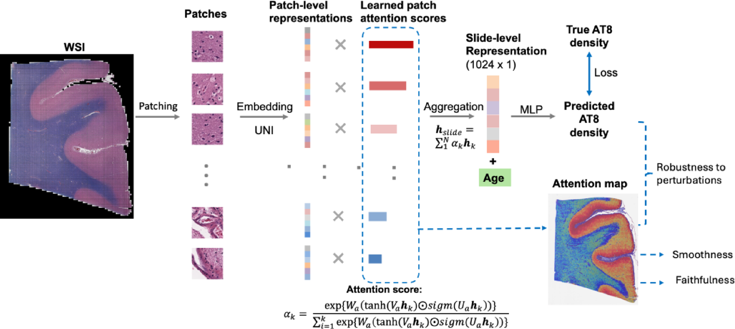
Age-informed, attention-based weakly supervised learning for neuropathological image assessment
Shuying Li, Maxwell Malamut, Ann McKee, Jonathan D Cherry, Lei Tian
Brain Informatics 12 (1), 27
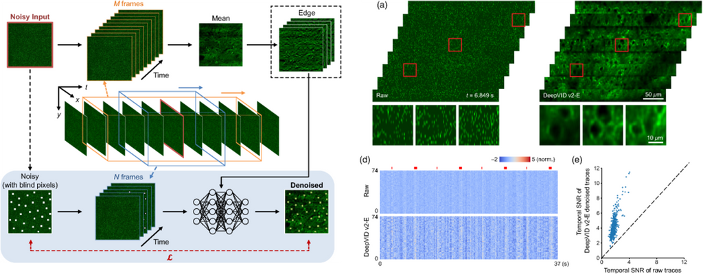
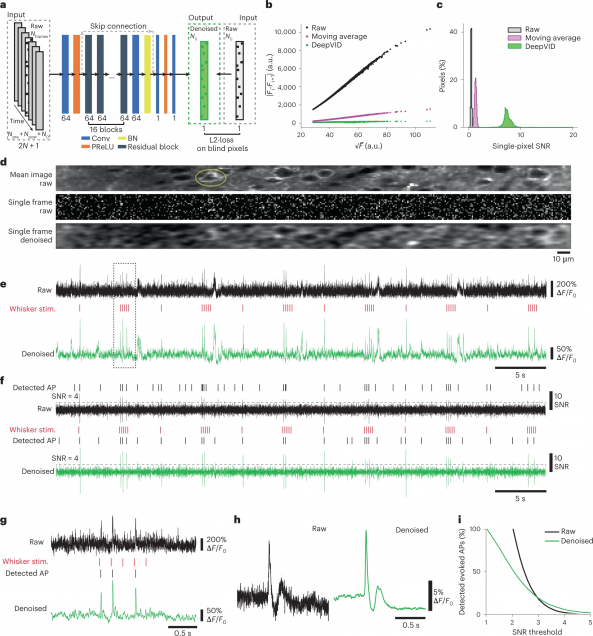
DeepVID v2: self-supervised denoising with decoupled spatiotemporal enhancement for low-photon voltage imaging
Chang Liu, Jiayu Lu, Yicun Wu, Xin Ye, Allison M. Ahrens, Jelena Platisa, Vincent A. Pieribone, Jerry L. Chen, Lei Tian
Neurophotonics, Vol. 11, Issue 4, 045007 (October 2024).
High-Speed Low-Light In Vivo Two-Photon Voltage Imaging of Large Neuronal Populations
Jelena Platisa, Xin Ye, Allison M Ahrens, Chang Liu, Ichun A Chen, Ian G Davison, Lei Tian, Vincent A Pieribone, Jerry L Chen
Nature Methods 20, 1095–1103 (2023).
⭑ Github Project
⭑ Spotlight: AI to the rescue of voltage imaging, Cell Reports Methods
Monitoring spiking activity across large neuronal populations at behaviorally relevant timescales is critical for understanding neural circuit function. Unlike calcium imaging, voltage imaging requires kilohertz sampling rates that reduce fluorescence detection to near shot-noise levels. High-photon flux excitation can overcome photon-limited shot noise, but photobleaching and photodamage restrict the number and duration of simultaneously imaged neurons. We investigated an alternative approach aimed at low two-photon flux, which is voltage imaging below the shot-noise limit. This framework involved developing positive-going voltage indicators with improved spike detection (SpikeyGi and SpikeyGi2); a two-photon microscope (‘SMURF’) for kilohertz frame rate imaging across a 0.4 mm × 0.4 mm field of view; and a self-supervised denoising algorithm (DeepVID) for inferring fluorescence from shot-noise-limited signals. Through these combined advances, we achieved simultaneous high-speed deep-tissue imaging of more than 100 densely labeled neurons over 1 hour in awake behaving mice. This demonstrates a scalable approach for voltage imaging across increasing neuronal populations.
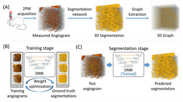
Anatomical modeling of brain vasculature in two-photon microscopy by generalizable deep learning
BME Frontiers, vol. 2021, Article ID 8620932
Segmentation of blood vessels from two-photon microscopy (2PM) angiograms of brains has important applications in hemodynamic analysis and disease diagnosis. Here we develop a generalizable deep-learning technique for accurate 2PM vascular segmentation of sizable regions in mouse brains acquired from multiple 2PM setups. In addition, the technique is computationally efficient, making it ideal for large-scale neurovascular analysis.
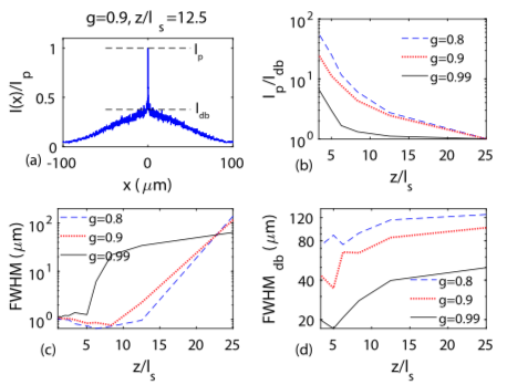
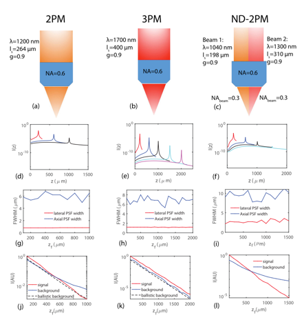
Comparing the fundamental imaging depth limit of two-photon, three-photon, and non-degenerate two-photon microscopy
Xiaojun Cheng, Sanaz Sadegh, Sharvari Zilpelwar, Anna Devor, Lei Tian, and David A. Boas
Opt. Lett. Vol. 45, Issue 10, pp. 2934-2937 (2020).
We have systematically characterized the degradation of imaging quality with depth in deep brain multi-photon microscopy, utilizing our recently developed numerical model that computes wave propagation in scattering media. The signal-to-background ratio (SBR) and the resolution determined by the width of the point spread function are obtained as functions of depth. We compare the imaging quality of two-photon (2PM), three-photon (3PM), and non-degenerate two-photon microscopy (ND-2PM) for mouse brain imaging. We show that the imaging depth of 2PM and ND-2PM are fundamentally limited by the SBR, while the SBR remains approximately invariant with imaging depth for 3PM. Instead, the imaging depth of 3PM is limited by the degradation of the resolution, if there is sufficient laser power to maintain the signal level at large depth. The roles of the concentration of dye molecules, the numerical aperture of the input light, the anisotropy factor , noise level, input laser power, and the effect of temporal broadening are also discussed.
Development of a beam propagation method to simulate the point spread function degradation in scattering media
Xiaojun Cheng, Yunzhe Li, Jerome Mertz, Sava Sakadžić, Anna Devor, David A. Boas, Lei Tian
Opt. Lett. 44, 4989-4992 (2019).
Scattering is one of the main issues that limit the imaging depth in deep tissue optical imaging. To characterize the role of scattering, we have developed a forward model based on the beam propagation method and established the link between the macroscopic optical properties of the media and the statistical parameters of the phase masks applied to the wavefront. Using this model, we have analyzed the degradation of the point-spread function of the illumination beam in the transition regime from ballistic to diffusive light transport. Our method provides a wave-optic simulation toolkit to analyze the effects of scattering on image quality degradation in scanning microscopy.