Phase Retrieval
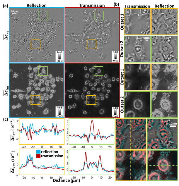
Inverse scattering for reflection intensity phase microscopy
Alex Matlock, Anne Sentenac, Patrick C. Chaumet, Ji Yi, and Lei Tian
Biomedical Optics Express Vol. 11, Issue 2, pp. 911-926 (2020).
Reflection phase imaging provides label-free, high-resolution characterization of biological samples, typically using interferometric-based techniques. Here, we investigate reflection phase microscopy from intensity-only measurements under diverse illumination. We evaluate the forward and inverse scattering model based on the first Born approximation for imaging scattering objects above a glass slide. Under this design, the measured field combines linear forward-scattering and height-dependent nonlinear back-scattering from the object that complicates object phase recovery. Using only the forward-scattering, we derive a linear inverse scattering model and evaluate this model’s validity range in simulation and experiment using a standard reflection microscope modified with a programmable light source. Our method provides enhanced contrast of thin, weakly scattering samples that complement transmission techniques. This model provides a promising development for creating simplified intensity-based reflection quantitative phase imaging systems easily adoptable for biological research.
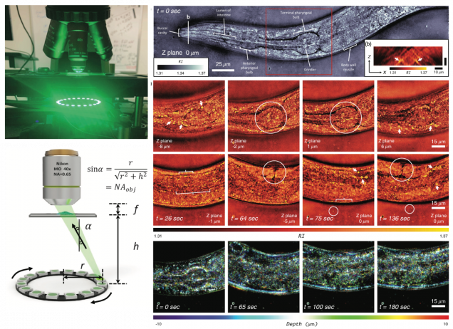
High-speed in vitro intensity diffraction tomography
Jiaji Li, Alex Matlock, Yunzhe Li, Qian Chen, Chao Zuo, Lei Tian
Advanced Photonics, 1(6), 066004 (2019).
⭑ Highlighted at Programmable LED ring enables label-free 3D tomography for conventional microscopes
We demonstrate a label-free, scan-free intensity diffraction tomography technique utilizing annular illumination (aIDT) to rapidly characterize large-volume 3D refractive index distributions in vitro. By optimally matching the illumination geometry to the microscope pupil, our technique reduces the data requirement by 60
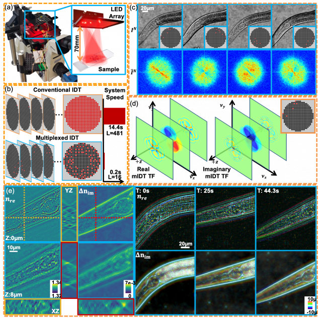
High-throughput, volumetric quantitative phase imaging with multiplexed intensity diffraction tomography
Alex Matlock, Lei Tian
Biomed. Opt. Express Vol. 10, Issue 12, pp. 6432-6448 (2019).
Intensity diffraction tomography (IDT) provides quantitative, volumetric refractive index reconstructions of unlabeled biological samples from intensity-only measurements. IDT is scanless and easily implemented in standard optical microscopes using an LED array but suffers from large data requirements and slow acquisition speeds. Here, we develop multiplexed IDT (mIDT), a coded illumination framework providing high volume-rate IDT for evaluating dynamic biological samples. mIDT combines illuminations from an LED grid using physical model-based design choices to improve acquisition rates and reduce dataset size with minimal loss to resolution and reconstruction quality. We analyze the optimal design scheme with our mIDT framework in simulation using the reconstruction error compared to conventional IDT and theoretical acquisition speed. With the optimally determined mIDT scheme, we achieve hardware-limited 4Hz acquisition rates enabling 3D refractive index distribution recovery on live Caenorhabditis elegans worms and embryos as well as epithelial buccal cells. Our mIDT architecture provides a 60 × speed improvement over conventional IDT and is robust across different illumination hardware designs, making it an easily adoptable imaging tool for volumetrically quantifying biological samples in their natural state.
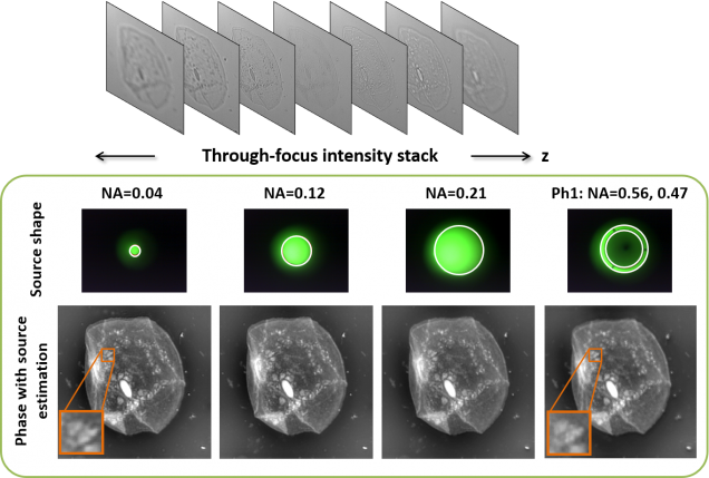
Nonlinear Optimization Algorithm for Partially Coherent Phase Retrieval and Source Recovery
J. Zhong, L. Tian, P. Varma, L. Waller
IEEE Transactions on Computational Imaging 2 (3), 310 – 322 (2016).
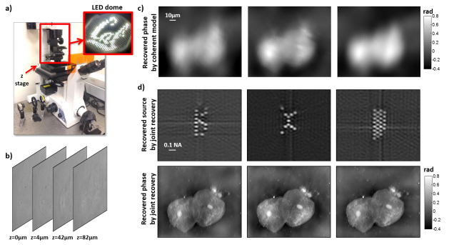
We propose a new algorithm for recovering both complex field (phase and amplitude) and source distribution (illumination spatial coherence) from a stack of intensity images captured through focus. The joint recovery is formulated as a nonlinear least-square-error optimization problem, which is solved iteratively by a modified Gauss-Newton method. We derive the gradient and Hessian of the cost function and show that our second-order optimization approach outperforms previously proposed phase retrieval algorithms, for datasets taken with both coherent and partially coherent illumination. The method is validated experimentally in a commercial microscope with both Kohler illumination and a programmable LED dome.
Experimental robustness of Fourier Ptychography phase retrieval algorithms
L. Yeh, J. Dong, J. Zhong, L. Tian, M. Chen, G. Tang, M. Soltanolkotabi, L. Waller
Opt. Express 23(26) 33212-33238 (2015).
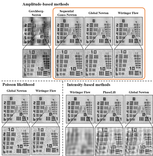
Fourier ptychography is a new computational microscopy technique that provides gigapixel-scale intensity and phase images with both wide field-of-view and high resolution. By capturing a stack of low-resolution images under different illumination angles, an inverse algorithm can be used to computationally reconstruct the high-resolution complex field. Here, we compare and classify multiple proposed inverse algorithms in terms of experimental robustness. We find that the main sources of error are noise, aberrations and mis-calibration (i.e. model mis-match). Using simulations and experiments, we demonstrate that the choice of cost function plays a critical role, with amplitude-based cost functions performing better than intensity-based ones. The reason for this is that Fourier ptychography datasets consist of images from both brightfield and darkfield illumination, representing a large range of measured intensities. Both noise (e.g. Poisson noise) and model mis-match errors are shown to scale with intensity. Hence, algorithms that use an appropriate cost function will be more tolerant to both noise and model mis-match. Given these insights, we propose a global Newton’s method algorithm which is robust and accurate. Finally, we discuss the impact of procedures for algorithmic correction of aberrations and mis-calibration.
3D intensity and phase imaging from light field measurements in an LED array microscope
Lei Tian, L. Waller
Optica 2, 104-111 (2015).
⭑ the 15 Most Cited Articles in Optica published in 2015 (Source: OSA, 2019)
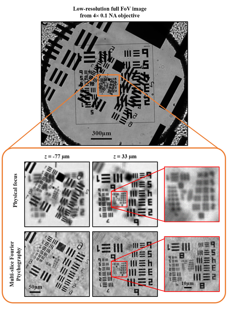
Realizing high resolution across large volumes is challenging for 3D imaging techniques with high-speed acquisition. Here, we describe a new method for 3D intensity and phase recovery from 4D light field measurements, achieving enhanced resolution via Fourier Ptychography. Starting from geometric optics light field refocusing, we incorporate phase retrieval and correct diffraction artifacts. Further, we incorporate dark-field images to achieve lateral resolution beyond the diffraction limit of the objective (5x larger NA) and axial resolution better than the depth of field, using a low magnification objective with a large field of view. Our iterative reconstruction algorithm uses a multi-slice coherent model to estimate the 3D complex transmittance function of the sample at multiple depths, without any weak or single-scattering approximations. Data is captured by an LED array microscope with computational illumination, which enables rapid scanning of angles for fast acquisition. We demonstrate the method with thick biological samples in a modified commercial microscope, indicating the technique’s versatility for a wide range of applications.
Partially coherent phase imaging with unknown source shape
J. Zhong, Lei Tian, J. Dauwels, L. Waller
Biomedical Optics Express 6, 257-265 (2015).
We propose a new method for phase retrieval that uses partially coherent illumination created by any arbitrary source shape in Kohler geometry. Using a stack of defocused intensity images, we recover not only the phase and amplitude of the sample, but also an estimate of the unknown source shape, which describes the spatial coherence of the illumination. Our algorithm uses a Kalman filtering approach which is fast, accurate and robust to noise. The method is experimentally simple and flexible, so should find use in optical, electron, X-ray and other phase imaging systems which employ partially coherent light. We provide an experimental demonstration in an optical microscope with various condenser apertures.
