Functional Neuroimaging of Speech
In order to test and refine our modeling framework, our lab designs and carries out a variety of brain imaging studies in humans. Most of our work has concentrated on the use of functional magnetic resonance imaging (fMRI), a technique that is sensitive to changes in blood oxygenation levels that result from local brain activations. This method thus allows us to obtain statistical maps of cortical activations that can easily be compared with simulations from our modeling work. Because speech is only available for study in humans, fMRI is an invaluable technique, as it is very safe and completely non-invasive. We also use diffusion-weighted imaging to investigate white matter pathways in the speech network of neurologically normal individuals and those with communication disorders.
Our lab has also developed a suite of region-of-interest (ROI) based analysis tools for fMRI that are available from our Software page. We have shown that analyzing imaging results based on anatomically defined regions of interest across subject, rather than using spatial averaging, can significantly enhance statistical power.
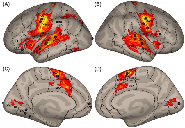
Brain activity while producing single words or pseudowords plotted on inflated cortical surfaces (adapted from Guenther, 2016, Chapter 2).
Publications
- Guenther, F.H. (2016). Neural Control of Speech. Cambridge, MA: MIT Press.
- Sitek, K.R., Cai, S., Beal, D.S., Perkell, J.S., Guenther, F.H., and Ghosh, S.S. (2016). Decreased cerebellar-orbitofrontal connectivity correlates with stuttering severity: Whole-brain functional and structural connectivity associations with persistent developmental stuttering. Frontiers in Human Neuroscience, 10, article 190.
- Segawa, J.A., Tourville, J.A., Beal, D.S., and Guenther, F.H. (2015). The neural correlates of speech motor sequence learning. Journal of Cognitive Neuroscience, 27, pp. 819-831. PMCID: PMC4344924
- Cai, S., Tourville, J.A., Beal, D.S., Perkell, J.S., Guenther, F.H. and Ghosh, S.S. (2014). Diffusion imaging of cerebral white matter in persons who stutter: evidence for network-level anomalies. Frontiers in Human Neuroscience, 8, pp. 54.
- Niziolek, C. and Guenther, F.H. (2013). Vowel category boundaries enhance cortical and behavioral responses to speech feedback alterations. Journal of Neuroscience, 33, pp. 12090-12098. PMCID: PMC3713738
- Golfinopoulos E., Tourville J.A., Bohland J.W., Ghosh S.S., Guenther F.H. (2011). fMRI investigation of unexpected somatosensory feedback perturbation during speech. NeuroImage 55, pp.1324-1338. PMCID:PMC3065208
- Golfinopoulos E., Tourville J.A., Guenther F.H. (2010). The integration of large-scale neural network modeling and functional brain imaging in speech motor control. NeuroImage 52, pp. 862-874. PMCID:PMC2891349
- Peeva, M.G., Guenther, F.H., Tourville, J.A., Neito-Castanon, A., Anton, J.L., Nazarian, B. and Alario, F.X. (2010). Distinct representations of phonemes, syllables, and supra-syllabic sequences in the speech production network. Neuroimage. 50, pp. 626-638. PMCID:PMC2840383
- Ghosh S.S., Tourville, J.A., and Guenther F.H. (2009) A neuroimaging study of premotor lateralization and cerebellar involvement in the production of phonemes and syllables. Journal of Speech, Language, and Hearing Research, 51(5), pp. 1183-1202. PMCID:PMC26522040
- Tourville, J.A., Reilly, K.J., and Guenther, F.H. (2008) Neural mechanisms underlying auditory feedback control of speech. NeuroImage, 39(3), pp. 1429-1443. PMCID:PMC3658624
- Bohland, J.W., and Guenther, F.H. (2006) An fMRI investigation of syllable sequence production. NeuroImage, 32(2), pp. 821-841. PMID:16730195
- Guenther, F.H., Nieto-Castanon, A., Ghosh, S.S., and Tourville, J.A. (2004) Representation of sound categories in auditory cortical maps. Journal of Speech, Language, and Hearing Research, 47(1), pp.46-57. PMID:15072527
- Nieto-Castanon, A., Ghosh, S.S., Tourville, J.A., and Guenther, F.H. (2003) Region-of-interest based analysis of functional imaging data. NeuroImage, 19, pp. 1303-1316. PMID:12948689
- Guenther, F.H., and Bohland, J.W. (2002) Learning sound categories: A neural model and supporting experiments. Acoustical Science and Technology, 23(4), pp. 213-220.