Chemical Imaging innovations
A central theme of our research is developing new ways to peer deeper into biology—methods that reveal the inner workings of cells and tissues without relying on dyes. Traditionally, scientists have visualized molecular processes through labeling techniques that use fluorescent dyes. These dyes, such as GFP, attach to specific molecules or cellular structures and emit light under a microscope, providing powerful tools for studying biological systems. However, despite their immense contributions to life sciences, fluorescent labels have fundamental limitations. On the molecular scale, these labels are often larger than the structures they bind to, obscuring precise details such as location, structure, and composition. In addition, the toxicity of the dyes and labeling processes prevents their direct application in human patients.
To overcome these limitations, our research leverages intrinsic molecular spectroscopic signals to visualize biological structures and dynamics in a completely label-free manner (Nature Methods, May 2025 Focus Issue on Bond-selective Imaging). Over the past two decades, our team has developed a suite of chemical imaging platforms that span the full spectrum of molecular spectroscopy—from transient absorption microscopy in the UV-visible region, to coherent Raman scattering microscopy in the near-infrared, overtone photoacoustic microscopy in the shortwave infrared, and mid-infrared photothermal microscopy in the mid-infrared region.
The three pillars of our innovation are
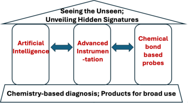
1.1 Pushing physical limits via advanced instrumentation
Our team constantly pushes the physical limits of chemical microscopy in terms of detection sensitivity, spatial resolution, imaging depth and speed. Major inventions are
• vibrational photoacoustic microscopy (Phys Rev Lett, 2011). Through photoacoustic detection of overtone vibration, we pushed the imaging depth from 100 micron in CARS to 6 millimeters.
• Saturated transient absorption microscopy (Nature Photonics 2013) as the first report of label-free STED using ground state depletion as mechanism.
• Mid-infrared photothermal (MIP) microscopy (Science Adv 2016) allows super-resolution infrared spectroscopic imaging of living systems.
• Stimulated Raman photothermal (SRP) microscopy (Science Adv 2023) boosts the detection sensitivity by 20 folds over SRS microscopy.
• Shortwave infrared photothermal (SWIP) microscopy (Nature Photonics 2024) allows millimeter-depth imaging of intracellular molecules.
• FURNACE SRS (Nature Methods 2025) pushes the resolution limit to 86 nm for intracellular metabolic imaging.
Funding:
- R35 GM136223, 2020 to 2029, Unveiling Hidden Signatures in Life by Vibrational Photothermal Microscopy, PI (Cheng)
- R01 EB035429, 2024 to 2027, Super-sensitive vibrational imaging by synergic development of instruments and Probes, PI (Cheng)
- R44 GM154516, 2024 to 2026, Video Rate Photothermal Infrared Spectroscopy, PI (Craig Prater)
- Chan Zuckerberg Initiative, 2023 to 2026, Bond-selective intensity diffraction tomography, PI (Cheng)
- BU Ignition Award, 2025 to2026, IR AMES for bio-nanoparticle analysis.
1.2 Chemical imaging with artificial intelligence
Our team integrates deep learning frameworks into chemical microscopy to break the tradeoff in the instrument’s parameter space. We have developed a series of deep learning methods for denoise a vibrational hyperspectral stack. Very recently, we started to embed AI into the hardware towards smart chemical imaging.
- Supervised deep learning denoising (Nature Communications 2021)
- Self-supervised denoising via NACE (Nature Methods 2025)
- Self-supervised denoising via SPEND (Cell Press, Newton, 2025)
Funding:
- R01 EB032391, 2022.1 to 2025.12, High-content High-speed Chemical Imaging of Metabolic Reprogramming by Integration of Advanced Instrumentation and Data Science, PI Cheng.
1.3 Chemical-bond based probes for bio-orthogonal chemical imaging
Our team develops and employs chemical bond-based probes for bio-orthogonal chemical imaging in the spectrally silent window (2000 to 2300 wavenumbers). Examples include stable isotope probes (C-D), azido photothermal probes, nitrile chameleon, and more recently, genetically coded vibrational proteins.
- Azido photothermal probe (App) as GPS of small molecules (Science Adv 2024)
- Nitrile chameleon for mapping of enzymatic activities in live cells (Nature Methods 2024)
Funding:
- R33 CA287046, 2024.9 to 2027.8, Bio-orthogonal mid-Infrared photothermal imaging of cancer metabolism, PI Cheng