Artificial Intelligence for Medical Imaging & MRI
We develop physics-informed AI frameworks that enable reliable MRI reconstruction and analysis from limited and accelerated data. By integrating domain knowledge with modern machine learning, these approaches achieve robust, transferable performance across imaging conditions and clinical tasks, advancing scalable, multi-task medical AI for real-world clinical deployment.

Magnetic resonance imaging (MRI) is a cornerstone of modern medical diagnosis, yet its broader clinical impact remains constrained by fundamental limitations—long acquisition times, high operational costs, and a strong dependence on specialized infrastructure. Addressing these challenges requires not only advances in hardware, but also computational paradigms that can extract more information from less data while maintaining clinical reliability. Our research follows a coherent trajectory toward this goal, evolving from physics-informed reconstruction to generalizable AI systems capable of operating under real-world constraints.
Accelerated MRI Reconstruction: From Data Scarcity to Reliable Imaging: 1, 2, 3 We begin by addressing one of the most immediate bottlenecks in MRI: slow acquisition and incomplete data. Accelerated MRI relies on undersampling in k-space, which significantly reduces scan time but introduces artifacts and information loss. To overcome this, we develop reconstruction methods that integrate deep learning with MRI physics, including hybrid architectures that jointly model k-space and image-domain information, as well as Bayesian approaches that leverage statistical priors to guide reconstruction. Our work demonstrates that high-quality images can be recovered even under aggressive undersampling while preserving structures critical for diagnosis. Importantly, we also investigate robustness under degraded conditions, particularly for clinically relevant tasks such as stroke detection. By incorporating distributionally robust learning strategies, our models maintain reliable diagnostic performance even when image quality is compromised, underscoring the importance of coupling reconstruction with downstream clinical objectives rather than treating them as independent problems.
Data-Efficient and Transferable Learning: 4, 5 Building on these reconstruction advances, we turn to a broader challenge: limited and heterogeneous medical data. Many clinical scenarios lack sufficient labeled datasets, and imaging conditions vary widely across scanners, institutions, and patient populations. To address this, we develop data-efficient learning frameworks that reduce reliance on large, task-specific annotations. Diffusion-based reconstruction models enable high-quality image recovery through novel degradation processes in k-space, while neural style transfer methods enable field-strength translation, generating high-quality images from low-field inputs without requiring paired training data. These approaches emphasize transferability and flexibility, demonstrating that models can generalize across imaging domains, anatomical regions, and acquisition conditions. This reflects a shift from optimizing for individual datasets to designing systems that remain robust under real-world variability.
Toward Unified and Generalizable Models: 6 A central question that emerges from this progression is whether MRI models can move beyond task-specific designs toward unified architectures. To this end, we introduce transformer-based frameworks such as the Magnetic Resonance Image Processing Transformer (MR-IPT), pretrained on diverse MRI datasets and applied across multiple reconstruction settings. By leveraging a shared backbone, these models learn universal feature representations that generalize across acceleration factors, sampling patterns, and perturbations. This marks a conceptual transition from task-specific learning to representation learning, where a single model can adapt to multiple scenarios without retraining from scratch. The result is enhanced robustness, scalability, and efficiency compared to conventional deep learning approaches.
From Reconstruction to Multi-Task Medical AI: 7 While reconstruction addresses image quality, clinical practice requires models that can also interpret and analyze images across tasks. A key limitation of earlier approaches is that generalization is often confined to a single task domain. To overcome this, our recent work explores whether pretrained models can extend across multiple downstream medical imaging tasks, even under limited labeled data. Using large-scale self-supervised pretraining on over 31 million MRI slices, we develop a unified transformer framework that supports MRI sequence classification, skull stripping (brain extraction), and multi-region anatomical segmentation. These models achieve strong performance in few-shot settings, demonstrating that large-scale pretraining can significantly reduce the need for extensive annotations. This progression reflects a broader shift—from reconstructing images to understanding them, and ultimately to enabling flexible, task-agnostic AI systems.
Integration of Physics, Learning, and Clinical Validation: Across all stages of this work, a unifying theme is the integration of physical modeling, statistical reasoning, and modern machine learning. Rather than treating MRI as a purely data-driven problem, we embed domain knowledge to ensure that models remain interpretable, stable, and firmly grounded in imaging principles. Equally important is clinical validation: our work is evaluated not only through quantitative metrics, but also in clinically relevant settings, including stroke diagnosis and cross-dataset generalization. Through this integration, methodological advances are translated into robust, clinically meaningful improvements, bridging the gap between algorithmic innovation and real-world medical practice.
Outlook: Toward Scalable and Practical Medical AI: The work highlighted here traces a clear progression from accelerating MRI acquisition to developing generalizable reconstruction models, and ultimately to enabling scalable, deployable AI systems for medical imaging. Looking ahead, we seek to advance these frameworks toward multimodal imaging that integrates MRI with complementary modalities such as CT and PET; toward disease-focused applications including dementia and neurodegenerative disorders; and toward clinical translation through sustained collaboration with radiologists and healthcare systems. By uniting advances in machine learning with domain knowledge and clinical insight, our vision is to bridge the gap between AI innovation and medical practice—enabling imaging systems that are not only reliable and scalable, but also broadly accessible, adaptive, and impactful across diverse clinical settings.
| Representative Publications (# denotes supervised by X. Zhang; * denotes corresponding author: X. Zhang) |
|
NeuroBridge: Bridging multi-task MRI knowledge for neurodegenerative disease diagnosis
M. Li#, G. Shen#, C.W. Farris, X. Zhang* arXiv, 2026, preprint arXiv:2607.01401 +AbstractINTRODUCTION: Accurate MRI-based identification of Alzheimer’s disease (AD), mild cognitive impairment (MCI), and related dementias remains challenging because disease-related structural changes are often subtle and heterogeneous. We developed NeuroBridge, a clinically guided multi-task MRI framework for neurodegenerative disease diagnosis. METHODS: NeuroBridge integrates large-scale self-supervised MRI pretraining with hippocampal segmentation, hippocampal atrophy classification, and reconstruction objectives, followed by gated fusion fine-tuning. Performance was evaluated across ADNI and OASIS cohorts, including cross-cohort transfer, probability-based analysis, and opportunistic screening. RESULTS: NeuroBridge achieved the highest performance across evaluated classification tasks, reaching 88.17% accuracy for AD versus cognitively normal controls in ADNI and 82.78% in OASIS. The largest gains occurred in MCI-related and mixed-diagnosis settings. The framework demonstrated strong cross-cohort generalization, systematic associations between predicted-class probability and accuracy, and the feasibility of probability-based opportunistic screening. DISCUSSION: Clinically guided multi-task representation learning improves neurodegenerative MRI diagnosis beyond conventional single-task approaches. NeuroBridge provides a robust and scalable framework for dementia assessment and MRI-based opportunistic screening.
|
|
Few-shot deployment of pretrained MRI transformers in brain imaging tasks
M. Li#, G. Shen#, C.W. Farris, and X. Zhang* Frontiers in Artificial Intelligence, 2026, 9: 1771088 +AbstractIntroduction: Transformer-based deep learning has shown great potential in medical imaging, but its real-world applicability remains limited due to the scarcity of annotated data. This study aims to develop a practical framework for the few-shot deployment of pretrained MRI transformers across diverse brain imaging tasks. Methods: We employ a Masked Autoencoder (MAE) pretraining strategy on a large-scale, multi-cohort brain MRI dataset comprising over 31 million 2D slices to learn transferable representations. For classification tasks, a frozen MAE encoder with a lightweight linear head (MAE-classify) is used. For segmentation, we propose MAE-FUnet, a hybrid architecture that fuses pretrained MAE embeddings with multi-scale CNN features. Extensive evaluations are conducted on multiple datasets, including NACC, ADNI, OASIS, NFBS, SynthStrip, and MRBrainS18, under controlled few-shot settings. Results: The proposed framework achieves state-of-the-art performance in MRI sequence classification, reaching an accuracy of 99.24% with only 6,152 trainable parameters. For segmentation tasks, MAE-FUnet consistently outperforms strong baselines, achieving superior Dice and IoU scores across skull stripping and multi-class anatomical segmentation benchmarks. The model also demonstrates enhanced robustness and stability under data-limited conditions, with lower performance variance compared to competing methods. Discussion: These results highlight the effectiveness of pretrained MAE representations for few-shot medical imaging tasks. The proposed framework enables efficient, scalable, and adaptable deployment of transformer-based models in data-constrained clinical environments. The fusion of global transformer embeddings with local CNN features provides a generalizable design paradigm for a wide range of medical imaging applications.
|
|
Magnetic resonance image processing transformer for general accelerated image restoration
G. Shen#, M. Li#, S.W. Anderson, C.W. Farris, and X. Zhang* Scientific Reports — Nature, 2025, 15: 40064 +AbstractRecent advancements in deep learning have enabled the development of generalizable models that achieve state-of-the-art performance across various imaging tasks. Vision Transformer (ViT)-based architectures, in particular, have demonstrated strong feature extraction capabilities when pre-trained on large-scale datasets. In this work, we introduce the Magnetic Resonance Image Processing Transformer (MR-IPT), a ViT-based image-domain framework designed to enhance the generalizability and robustness of accelerated MRI restoration. Unlike conventional deep learning models that require separate training for different acceleration factors, MR-IPT is pre-trained on a large-scale dataset encompassing multiple undersampling patterns and acceleration settings, enabling a unified framework. By leveraging a shared transformer backbone, MR-IPT effectively learns universal feature representations, allowing it to generalize across diverse restoration tasks. Extensive experiments demonstrate that MR-IPT outperforms both CNN-based and existing transformer-based methods, achieving superior quality across varying acceleration factors and sampling masks. Moreover, MR-IPT exhibits strong robustness, maintaining high performance even under unseen acquisition setups, highlighting its potential as a scalable and efficient solution for accelerated MRI. Our findings suggest that transformer-based general models can significantly advance MRI restoration, offering improved adaptability and stability compared to traditional deep learning approaches.
|
|
Regularization by neural style transfer for MRI field-transfer reconstruction with limited data
G. Shen#, Y. Zhu, M. Li#, R. McNaughton#, H. Jara, S.B. Andersson, C.W. Farris, S.W. Anderson, and X. Zhang* Frontiers in Artificial Intelligence, 2025, 8: 1579251 +AbstractRecent advances in MRI reconstruction have demonstrated remarkable success through deep learning-based models. However, most existing methods rely heavily on large-scale, task-specific datasets, making reconstruction in data-limited settings a critical yet underexplored challenge. While regularization by denoising (RED) leverages denoisers as priors for reconstruction, we propose Regularization by Neural Style Transfer (RNST), a novel framework that integrates a neural style transfer (NST) engine with a denoiser to enable magnetic field-transfer reconstruction. RNST generates high-field-quality images from low-field inputs without requiring paired training data, leveraging style priors to address limited-data settings. Our experiment results demonstrate RNST’s ability to reconstruct high-quality images across diverse anatomical planes (axial, coronal, sagittal) and noise levels, achieving superior clarity, contrast, and structural fidelity compared to lower-field references. Crucially, RNST maintains robustness even when style and content images lack exact alignment, broadening its applicability in clinical environments where precise reference matches are unavailable. By combining the strengths of NST and denoising, RNST offers a scalable, data-efficient solution for MRI field-transfer reconstruction, demonstrating significant potential for resource-limited settings.
|
|
Learning to reconstruct accelerated MRI through K-space cold diffusion without noise
G. Shen#, M. Li#, C.W. Farris, S.W. Anderson, and X. Zhang* Scientific Reports — Nature, 2024, 14: 21877 +AbstractDeep learning-based MRI reconstruction models have achieved superior performance these days. Most recently, diffusion models have shown remarkable performance in image generation, in-painting, super-resolution, image editing and more. As a generalized diffusion model, cold diffusion further broadens the scope and considers models built around arbitrary image transformations such as blurring, down-sampling, etc. In this paper, we propose a k-space cold diffusion model that performs image degradation and restoration in k-space without the need for Gaussian noise. We provide comparisons with multiple deep learning-based MRI reconstruction models and perform tests on a well-known large open-source MRI dataset. Our results show that this novel way of performing degradation can generate high-quality reconstruction images for accelerated MRI.
|
|
Attention hybrid variational net for accelerated MRI reconstruction
G. Shen#, B. Hao, M. Li#, C.W. Farris, I.C. Paschalidis, S.W. Anderson, and X. Zhang* APL Machine Learning, 2023, 1(4): 046116 +AbstractThe application of compressed sensing (CS)-enabled data reconstruction for accelerating magnetic resonance imaging (MRI) remains a challenging problem. This is due to the fact that the information lost in k-space from the acceleration mask makes it difficult to reconstruct an image similar to the quality of a fully sampled image. Multiple deep learning-based structures have been proposed for MRI reconstruction using CS, in both the k-space and image domains, and using unrolled optimization methods. However, the drawback of these structures is that they are not fully utilizing the information from both domains (k-space and image). Herein, we propose a deep learning-based attention hybrid variational network that performs learning in both the k-space and image domains. We evaluate our method on a well-known open-source MRI dataset (652 brain cases and 1172 knee cases) and a clinical MRI dataset of 243 patients diagnosed with strokes from our institution to demonstrate the performance of our network. Our model achieves an overall peak signal-to-noise ratio/structural similarity of 40.92 ± 0.29/0.9577 ± 0.0025 (fourfold) and 37.03 ± 0.25/0.9365 ± 0.0029 (eightfold) for the brain dataset, 31.09 ± 0.25/0.6901 ± 0.0094 (fourfold) and 29.49 ± 0.22/0.6197 ± 0.0106 (eightfold) for the knee dataset, and 36.32 ± 0.16/0.9199 ± 0.0029 (20-fold) and 33.70 ± 0.15/0.8882 ± 0.0035 (30-fold) for the stroke dataset. In addition to quantitative evaluation, we undertook a blinded comparison of image quality across networks performed by a subspecialty trained radiologist. Overall, we demonstrate that our network achieves a superior performance among others under multiple reconstruction tasks.
|
|
Bayesian reconstruction of magnetic resonance images using Gaussian processes
Y. Xu, C.W. Farris, S.W. Anderson, X. Zhang, and K.A. Brown Scientific Reports — Nature, 2023, 13: 12527 +AbstractA central goal of modern magnetic resonance imaging (MRI) is to reduce the time required to produce high-quality images. Efforts have included hardware and software innovations such as parallel imaging, compressed sensing, and deep learning-based reconstruction. Here, we propose and demonstrate a Bayesian method to build statistical libraries of magnetic resonance (MR) images in k-space and use these libraries to identify optimal subsampling paths and reconstruction processes. Specifically, we compute a multivariate normal distribution based upon Gaussian processes using a publicly available library of T1-weighted images of healthy brains. We combine this library with physics-informed envelope functions to only retain meaningful correlations in k-space. This covariance function is then used to select a series of ring-shaped subsampling paths using Bayesian optimization such that they optimally explore space while remaining practically realizable in commercial MRI systems. Combining optimized subsampling paths found for a range of images, we compute a generalized sampling path that, when used for novel images, produces superlative structural similarity and error in comparison to previously reported reconstruction processes (i.e. 96.3% structural similarity and < 0.003 normalized mean squared error from sampling only 12.5% of the k-space data). Finally, we use this reconstruction process on pathological data without retraining to show that reconstructed images are clinically useful for stroke identification. Since the model trained on images of healthy brains could be directly used for predictions in pathological brains without retraining, it shows the inherent transferability of this approach and opens doors to its widespread use.
|
|
Distributionally robust image classifiers for stroke diagnosis in accelerated MRI
B. Hao, G. Shen#, R. Chen, C.W. Farris, S.W. Anderson, X. Zhang, and I. Paschalidis MICCAI 2023, Springer, 14224: 768–777 +AbstractMagnetic Resonance Imaging (MRI) acceleration techniques using k-space sub-sampling (KS) can greatly improve the efficiency of MRI-based stroke diagnosis. Although Deep Neural Networks (DNN) have shown great potential on stroke lesion recognition tasks when the MR images are reconstructed from the full k-space, they are vulnerable to the lower quality MR images generated by KS. In this paper, we propose a Distributionally Robust Learning (DRL) approach to improve the performance of stroke recognition DNN models when the MR images are reconstructed from the sub-sampled k-space. For Convolutional Neural Network (CNN) and Vision Transformer (ViT)-based models, our methods improve the stroke classification AUROC and AUPRC by up to 11.91% and 9.32% on the KS-perturbed brain MR images, respectively, compared against Empirical Risk Minimization (ERM) and other baseline defensive methods. We further show that DRL models can successfully recognize the stroke cases from highly perturbed MR images where clinicians may fail, which provides a solution for improved diagnosis in an accelerated MRI setting.
|
| Ph.D. Dissertation |
| Towards generalizable deep learning for accelerated MRI reconstruction Guoyao Shen, Ph.D. Dissertation, Boston University (Advisor: Xin Zhang; June 2026) |
| Selected Media Articles |
Revolutionizing MRI Restoration with Transformer Technology “In an era marked by rapid advancements in medical imaging, researchers have unveiled a groundbreaking methodology that harnesses the power of transformer models in magnetic resonance imaging (MRI). This innovative approach promises to significantly enhance the process of image restoration, particularly for accelerated MRI scans, which are crucial for timely diagnoses in clinical settings. The implications of this research are profound, potentially transforming how clinicians acquire and interpret magnetic resonance images.” [Read More] |
Can We Train AI to Read an MRI?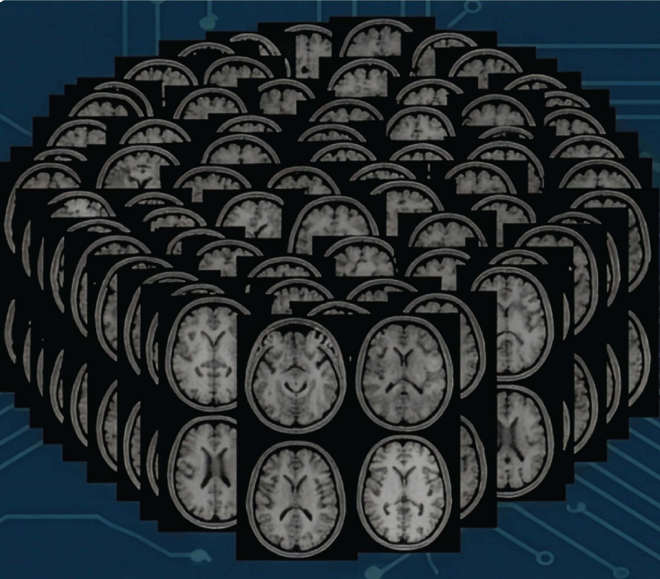 “When it comes to medical imaging, a chatbot doesn’t know its backside from its elbow. Or, at least, it can’t tell a spine from a knee. But if a Boston University research team is successful, a smarter AI system for evaluating brain scan images might someday help clinicians spot diseases sooner, saving lives while cutting costs.” [Read More] |