
Immunohistochemical staining of hnRNP H following KCl depolarization in rat primary neurons » Immunohistochemical staining of hnRNP H following KCl depolarization in rat primary neurons
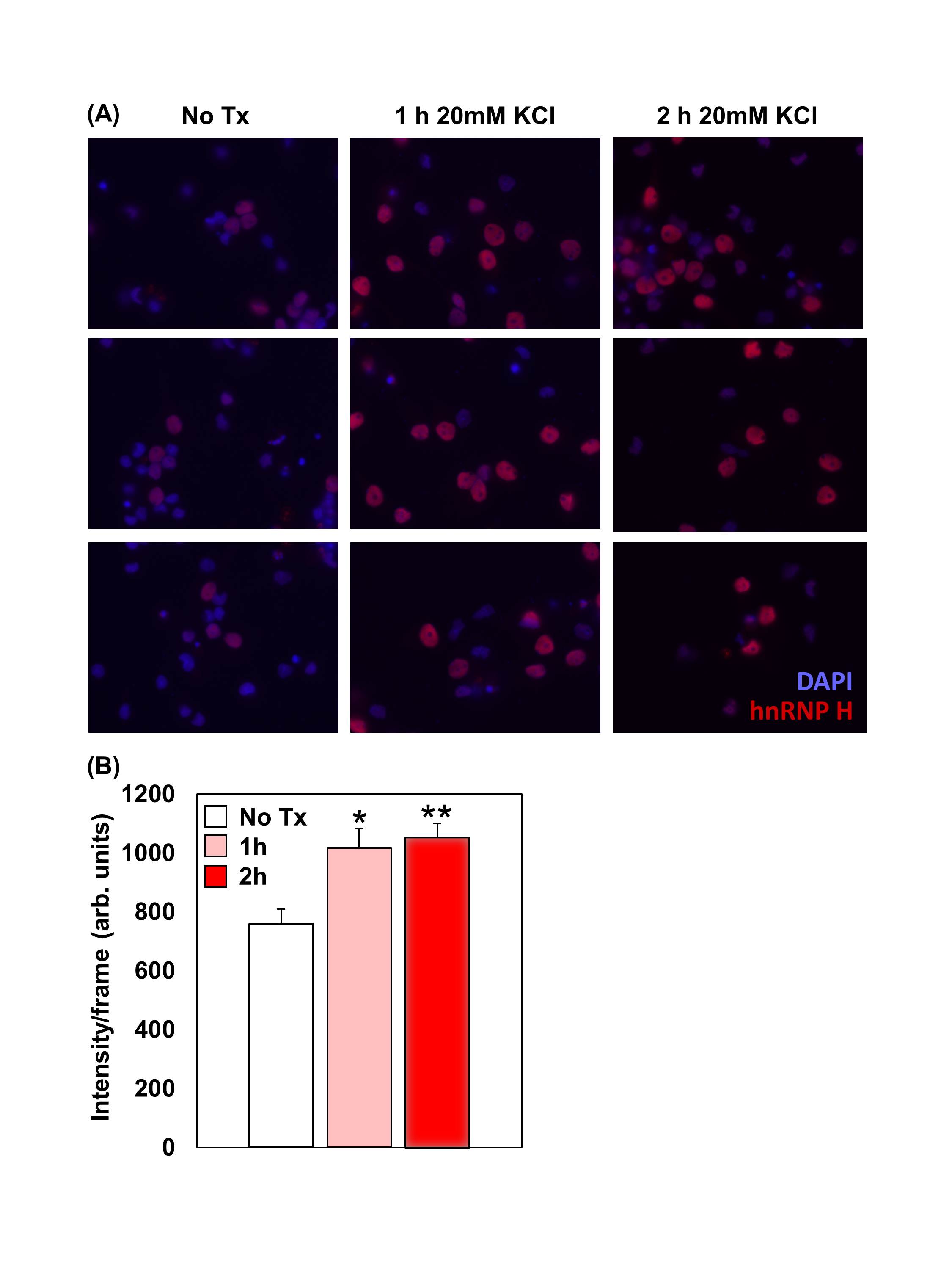
(A): Primary neocortical neurons were dissected from E18 Sprague-Dawley rat embryos (Charles River Laboratories). Dissociated neurons were cultured neurons for 1 week. For the control, no treatment (No Tx) group, 1 ml of conditioned media was replaced with 1 ml of neurobasal media. For the 1 h and 2 h Tx groups, 1 mL of conditioned media was replaced with 1ml of 20 mM KCl-enriched neurobasal media. Treated neurons were then washed, fixed, permeabilized, blocked, and incubated with primary hnRNP H antibody (1:500 Rabbit polyclonal, Bethyl Labs) in 1% BSA overnight at 4° C. 12 h later, neurons were washed and incubated with an Alexa Fluor 594 antibody (1:500 Donkey anti-Rabbit, Life Technologies) in 1% BSA. Processed coverslips were then stained with DAPI (blue) and mounted onto glass slides. Images were collected using a Zeiss AxioObserver microscope under uniform settings for all three groups. 20 serial images (frames) were captured per condition and fluorescence was quantified using ImageJ under a uniform threshold range. Note both an increase in the number of H1 stained neurons following 1-2 h of KCl Tx as well as an increase in the fluorescent staining intensity after KCl treatment. (B): Semi-quantification of fluorescence staining intensity. One-way ANOVA indicated a main effect of genotype (F2,57 = 8.4; P = 0.0006). *P = 0.01; **P <0.001 (unpaired t-tests versus No Tx).