Category: Article
The Effect of State and Trait Anxiety on Attention

A topic that has become more and more prevalent over the last few years is the effect of anxiety on attentiveness. How anxiety affects attention depends on the type of anxiety that is experienced. Most people experience state anxiety, in other words, a higher threat value is placed on a particular situation or stimulus. Feeling anxious during a very important midterm is an example of state anxiety. Another type of anxiety is trait anxiety, which is the tendency to focus one’s attention towards the stimulus causing the anxiety. In other words, to think only about the exam, instead of non-anxiety inducing stimuli.
Attention itself is composed of three independent networks: the alerting network, the orienting network, and the executive network. The alerting network is responsible for maintaining an appropriate level of sensitivity to the stimulus through activation of the right frontal and parietal areas of the brain. The orienting network attends to information coming from a specific stimulus among numerous sensory stimuli. This network is associated with activation in the superior parietal lobe, frontal eye fields, and temporoparietal junction. The executive network is responsible for conflict resolution and voluntary action control of the stimulus, and is related to the medial frontal areas of the brain, as well as the cingulate gyrus and lateral prefrontal cortex.
Two experiments were performed by the Department of Psychology and Physiology at the University of Granada and the Department of Neurology at Washington University School of Medicine to learn more about the relationship between these two different types of anxiety and attention. The first focused on trait anxiety, while the second focused on state anxiety.
The first experiment featured a group of individuals with either high or low trait anxiety, and another group of individuals with average trait anxiety. The two groups were made to perform various computer tasks that played to each specific network of attention. What the researchers found was that individuals with high-trait-anxiety had a more difficult time controlling interference in the computer tasks than those with low-trait-anxiety. However, the results of the alerting and orienting tasks were very similar for both groups. Those with high-trait anxiety had a difficult time responding to the task’s demands.
The second experiment also separated participants into two groups for anxious mood induction and non-anxious mood induction. One group was shown pleasant pictures and the other was shown unpleasant pictures, both groups were tasked with becoming emotionally involved in what they were seeing. In the group shown the negative stimuli (the unpleasant pictures), more emphasis was put on the lack of control over the negative circumstances represented in the image. The positive stimuli (the pleasant pictures) in the other group focused more on goal achievement. The individuals shown the negative stimuli revealed higher levels of anxiety than those shown the positive stimuli.
The results of the experiments revealed that both state and trait anxiety have a significant impact on attentional networks. State anxiety was found to have a greater impact on the alerting and orienting networks of attention, because they are more closely related to contextual sensitivity and vigilance processes. High state anxiety was found to be the result of a heightened response in the amygdala and superior temporal sulcus (regions activated during the assessment of valence facial expressions). It was also found that the executive network was less efficient in those with high-trait-anxiety than those with low-trait-anxiety because the executive network determines control, which is what those with high-trait-anxiety were lacking. High trait anxiety was related to a reduced prefrontal response (the region related to controlling complex processes). It was concluded that trait anxiety was responsible for a “reduced-general cognitive control capacity”.
-Allyson Pulsoni
Sources
1. Attention and Anxiety: Different Attentional Functioning Under State and Trait Anxiety
http://pss.sagepub.com.ezproxy.bu.edu/content/21/2/298.full.pdf+html
Image by Evan89, via Wikimedia Commons
Gene Regulation and The Hippocampus
Memory has always been and still is a subject of interest for many neuroscientists. Whether it pertains to increasing memory or finding cures to neurodegenerative diseases such as Alzheimer’s, memory is always a topic of interest for neuroscientists. Recently, there have been studies conducted by both the Institute of Basic Science and Advancing Science, Serving Society (AAAS) that have found genes that are suppressed or inhibited that prevent the formation of these long term memories. Increased hippocampal activity also is shown to have treatments toward age related memory loss, which could relate to the genes found by the AAAS and the IBS.
The IBS Center, using a technique called Ribosome profiling, was able to analyze the hippocampus of a mouse model and showed that gene regulation actually suppressed the formation of memory. According to the article “When an animal experiences no stimulus in an environment the hippocampus undergoes gene repression which prevents the formation of new memories”, showing activity of the neurons help prevent this gene regulation which allows the formation of memory. Natasha Pinol from the AAAS also found a similar action in his studies however showed that one estrogen receptor actually helped regulate the memories after learning instead of inhibiting it, as Jun Cho for IBS has found.
Although these particular genes themselves might still have their purposes unknown to the world, these findings can definitely be used to help with age related memory loss such as Alzheimer’s disease. Through a study done at Northwestern University by Marla Paul, it is shown that decreasing certain activity and increasing other activity could lead to an increase in memory formation. Although they did not target the same genes that Cho and Pinol targeted, this really shows how much more we are learning about the hippocampus, as memory is formed through a moderation of genes.
Sources:
http://neurosciencenews.com/memory-formation-gene-suppression-2799/
http://neurosciencenews.com/hippocampus-genetics-memory-formation-2807/
http://neurosciencenews.com/ca3-hippocampus-neurons-memory-aging-2709/
- Albert Wang
Is Music Affecting Our Memory?
 Music has been scientifically proven as beneficial, having effects such as reducing stress, enhancing blood vessel function, improving sleep quality, and improving cognitive performance. However, one thing that music does not improve is one’s ability to focus. In a recent study conducted at Georgia Institute of Technology, researchers found that listening to music decreased the efficiency of remembering names.
Participants in this study were asked to match faces to names, a task that involves associative memory. In associative memory, a memory of an event or place is triggered by the recollection of something associated with it. Music is heavily involved in associative memory, which is why it can be upsetting to listen to certain songs if you have associated them with an ex-significant other. Much like other types of memory, associative memories are processed in the hippocampus of the brain.
Some participants completed this name-face test in silence, while others had non-lyrical music playing in the background. All age groups of participants agreed that the music was distracting from the test, but only the scores of the older adults were affected by it.
Nanotechnology: Creating the perfect human?

How would you feel if you had the choice of having billions of tiny robots injected into your body? A pretty unpleasant thought, am I right? What if I told you that these tiny robots could repair any mutation you may have in your DNA? Sound far-fetched? Well, scientists have been making huge breakthroughs in this! It's called nanotechnology. These small robots are like tiny computers that are coded to attach to specific cells in your body and carry them from point A to point B. These tiny robots, 1-100nm in size (or 1 and 100 billionth of a meter!), are like transporters; they pick up the target cell at point A and move it to point B. Point B can be anything from the trash, (cell death) if the cell is not needed anymore, to another part of the body where the cell is needed. They also have the ability to reprogram a cell's biology. If more of one cell is needed in a particular area it can bring that cell to the specified area and "tell" it to replicate. Basically, nanotechnology will eventually perfect every single cell in your body.
The Mystery of the Human Connectome
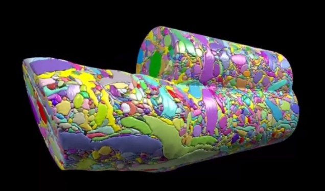
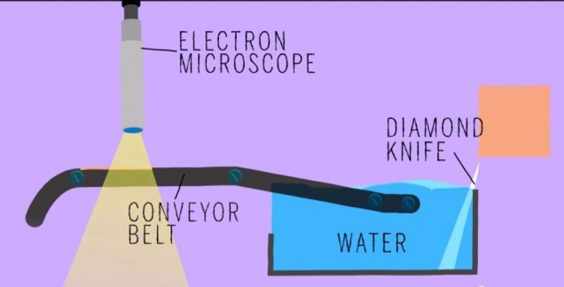
Did anyone read that article in BU today last week called “Untangling the Connectome?” In case you didn’t, here’s a brief synopsis. Dr. Kasthuri and his lab members are working on identifying the connections between the neurons in the human brain. This has been done before for C. elegans, which have only 302 neurons (compared to our 100 trillion). So, imagine how complex this project is and how much data is contained in one tiny slice of human brain?! In the article, Kasthuri said that a brain slice with the volume of “a millimeter cubed, at the resolution we would like, is about two million gigabytes of data” [1].That’s crazy. The Kasthuri lab implemented a clever method (Figure 1) to take images of 30 nm brain slices that provide the high resolution pictures they’re looking for.
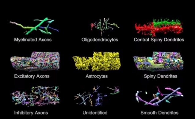
Basically, the brain volume is immersed in plastic and cut with a diamond knife to ensure precision. Since the slices are somewhat floppy, they are collected in water then picked up by a conveyor belt. While on the conveyor belt, an electron microscope takes a quick shot of the brain tissue and next a collection of images is put together to create a accurate image of the initial brain volume. Methods can be used to break down the image of the brain volume into components such as axons, dendrites, etc. (Figure 2). Pretty cool, right? The implications of this research is that understanding the connections in our brain can teach us about brain development and memory functions.
The Human Connectome project at Mass General Hospital (MGH) is working on developing high resolution neuroimaging methods. They’re working on figuring out how varying white matter fiber connectivity correlates to brain function and how cytoarchitectonics (the pattern of neuron organization/density in different areas of the brain, used to decipher Brodmann’s areas in the brain) can influence brain connectivity [2]. Methods they’re using include diffusion spectrum imaging (DSI), which is similar to diffusion tensor imaging (DTI) as both allow scientists to observe white matter connectivity since water diffuses parallel to white matter tracts. DTI measures the preferred and mean direction of the diffusion of water while DSI measures it in many different directions [3]. Scientists at MGH are also working on developing tools for high angular diffusion imaging (HARDI), which “address[es] the challenge of imaging crossing fibers by applying a stronger magnetic gradient for a longer time and capturing many more images of diffusion in various directions” [3].
Researchers are hoping to find patterns of connectivity by deciphering how our 100 trillion neurons are linked. This information could help scientists better understand the roots of different neurological or neurodegenerative diseases where perhaps neural connectivity differs significantly. This is a challenging project but the results of it could be a major breakthrough for neuroscience.
~ Srijesa K.
Sources:
[1] "Untangling the Connectome"
[3] "White's the Matter: A basic guide to white matter imaging using diffusion MRI"
Is iPhone Separation Anxiety Real?
 Have you ever searched for a ringing phone, feeling your anxiety increase with each ring? Or experienced a mini heart attack when you thought you lost your phone only to discover it was in a different pocket? Most older generations would criticize you for being so obsessed with technology, but recent studies have shown that ‘iPhone separation anxiety’ is a real disorder – and it is plaguing the younger generations of today’s society.
The average person spends about two hours and fifty-seven minutes on a smartphone or tablet every day. Most of us get stressed out if we misplace our phone, are constantly checking for notifications, and even feel ‘phantom vibrations’ – a sensation that we have received a notification when we really have not. Most people would dismiss this anxiety as an unhealthy obsession with technology, but research has shown that these feelings are legitimate and smartphone separation can have serious psychological and physiological effects.
Optogenetics: Using Light to Turn Memories On and Off

Scientists have known about rhodopsins that are responsible for sensing light for a while. What if there was a way to insert those rhodopsins inside neurons? That’s exactly what scientists were experimenting with in the early 2000s and it’s this idea that lead to the birth of optogenetics. By taking the DNA of channel rhodopsins from algae and inserting them into the membrane of neurons, scientists were able to make neurons sensitive to particular wavelengths of light. Channel rhodopsin and halorhodopsin are among the opins inserted into neurons by injecting viruses. Channel rhodopsin activates neurons while halorhodopsin silences them. Once the neuron expresses the light-gated cation channel channelrhodopsin-2 in its cells membrane, shining light on it for as little as a few milliseconds has a profound effect. It causes the opening of the channelrohodopsin-2 molecules, allowing positively charged ions to enter cell and cause the cell to fire.

Check out this video to see how optogenetics works.
Many experiments today use optogenetics to selectively turn neurons on and off in mice. What makes this method mind blowing is the high spatial and temporal resolution it gives scientists when working with the brain. It can be used on neurons in on a petri dish or within a living animal. It could be used to learn more about the function of particular brain regions. For instance, one could temporarily inactivate one region to observe how it impacts activity in other connected brain regions.
Furthermore, it’s minimally invasive: once the virus containing the rhodopsin has been injected, all the scientist needs to do is shine a pulse of light. Researchers at Stanford have used optogenetics to induce muscle contractions in mice. At Case Western Reserve University, researchers implemented it to restore motor function in rats paralyzed by spinal cord injuries. Could optogenetics be used to recover vision loss, something most humans deal with as they age? Experiments conducted on mice with a lack of photoreceptors shows that shining light on bipolar cells (containing channelrhodopsin-2) causes action potentials to fire in the visual cortex. It would be amazing if scientists could overcome biomedical and technical obstacles to make this work in humans too.
Across the river at MIT, members of the Tonegawa lab have taken the technique of optogenetics one step further. Steve Ramirez and Xu Liu have been working to localize memories in the brain and activate them with a light “switch”. And they have accomplished this feat in mice. Promising experiments with mice suggest optogenetics can be used to turn off traumatizing memories and activate pleasant once. This could have implications for PTSD, where horrific memories could be suppressed. They also have experimented with the idea of implanting false memories into the brain, which they call “Project Inception.” For more information on this work (and a good laugh), check out Steve Ramirez and Xu Liu’s TED talk.
Seems to me like optogenetics is a promising technique that can lead to breakthroughs in neuroscience.
-Srijesa K.
Sources
[1] “The Birth of Optogenetics” http://www.the-scientist.com/?articles.view/articleNo/30756/title/The-Birth-of-Optogenetics/
[2] “Potential Benefits of Optogenetics” http://optogenetics.weebly.com/what-is-it1.html
The Miracle of Neurogenesis
Neurogenesis occurs in two areas in the human adult: in the dentate gyrus of the hippocampus and in the olfactory system. The hippocampus is vital to learning new information and memory consolidation, thus it makes sense that new neurons need to be born in that region. The olfactory system is needs neurogenesis to process to new information. Majority of neurogenesis actually occurs during prenatal development. In fact humans initially have more neurons that necessary for survival. Apoptosis (programmed cell death) occurs to prune the synapses established during early development.
Many studies have been conducted that investigate ways to increase neurogenesis. Such activities include voluntary physical exercise or being in enriched environments. Experiments with rats have shown that being in an enriched environment where rats are exposed to complex objects, toys, running wheels, etc. spark improvements in performance of tasks measuring levels of learning and memory. For humans, I suppose an enriched environment could be a place involving novel stimuli or
If you think about it, how could neurogenesis be bad? I mean people with neurodegenerative diseases suffer from the consequences of neuronal loss, right? However, according to a study published in May 2014 in Science, exercise could induce amnesia. An article regarding this study states, “Adult mice that exercised on a running wheel after experiencing an event were more likely than their inactive mates to forget the experience.” Thus it appears that the neurogenesis that occurs during exercise may be “wiping out” neurons that encoded previous memories. Furthermore, when neurogenesis was pharmacologically inhibited scientists observed a recall failure in the rats. This article relates this phenomenon to the fact that children cannot form long term memories until they are 3-4 years of age.
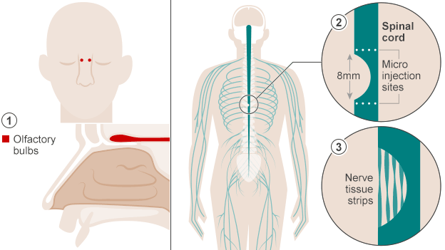
Despite controversy about neurogenesis, olfactory ensheathing cells (OECs) have been used to help a paralyzed man walk once again. An article published in October in BBC News describes how doctors in Poland accomplished this feat. The first step to this process was to extract cells from the patient’s olfactory bulbs (they removed one olfactory bulb and grew the cells in culture). Two weeks later the cells were implanted in the areas surrounding the spinal cord damage the patient had experienced. This action allowed the spinal cord cells to regenerate because the nerve grafts acted “as a bridge to cross the severed cord.” The implications of this type of surgery are pretty amazing – it could work wonders for paralyzed veterans/other individuals and people dealing with dysfunctions relevant to spinal cord damage.
Sources:
Exercise Can Erase Memories - The Scientist
Paralyzed Man Walks Again After Cell Transplant - BBC
- Srijesa K.
Is the brain the only place that stores our memories?
 Do you ever think about your childhood or replay an event in your head that happened 15 years ago but its so vivid that it seems like it happened yesterday? Do you ever hear something and think it sounds like your favorite song and then start singing that song? These are memories that were formed in your brain that are replayed as a result of a specific stimulus. For a long time scientists believed that memories were formed, processed, and sent to different destinations in the brain. Dr. Wilder Penfield was one of the first to accidentally discover this. In the 40s he electrically stimulated different areas of his patients' brains while they were under local anesthesia and found that the region he stimulated would elicit specific memories in the patient's life (see video below). For example, in one of his patients he stimulated her temporal lobe (auditory cortex) and she started to hum her favorite song out loud. This suggested that the memory of this song was stored in the place where it was processed or originated (i.e. the auditory cortex processed the first time she listened to the song). Penfield concluded that the cortex (the outer layers of the brain) stored the "complete record of the stream of consciousness; all those things in which a man was aware at any time..." Until recently, scientists have believed this phenomenon.
Stockholm Syndrome Explained by the Stanford Prison Experiment
Stockholm Syndrome can be referred to as a joke in the popular culture, and many people do not take it seriously as much as other common psychiatric problems such as PTSD, a psychological illness usually caused by a traumatic event like physical aggression. It cannot be treated seriously because there is no medical standard to properly diagnose a person with “Stockholm Syndrome.” However, this supposed illness is a real problem that affects the minority of people who are abducted usually by criminals who have no interest in the hostage’s safety.
The first recorded case of hostages with Stockholm Syndrome was during a bank robbery in Stockholm, Sweden. In August 23 to 28, 1973, the bank robbers negotiated with the police to leave the bank safely. While trying to form an agreement, the majority of the captive bank employees were unusually sympathetic towards the robber, and, even after being set free, refused to leave their captors. The criminologist and the psychiatrist who were investigating the robbery coined the term for their conditions “Stockholm Syndrome.”
