The Mystery of the Human Connectome
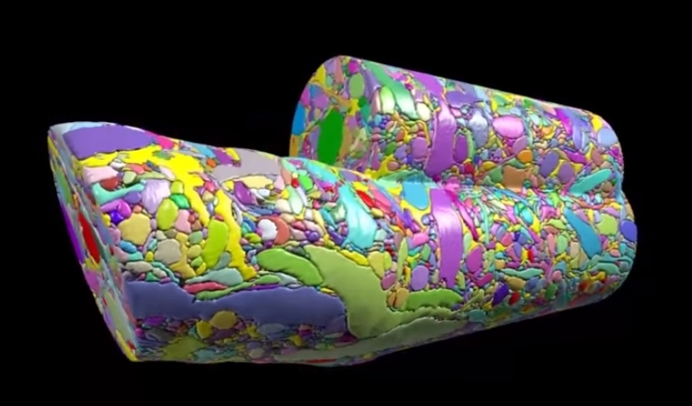
Section of brain volume.
Did anyone read that article in BU today last week called “Untangling the Connectome?” In case you didn’t, here’s a brief synopsis. Dr. Kasthuri and his lab members are working on identifying the connections between the neurons in the human brain. This has been done before for C. elegans, which have only 302 neurons (compared to our 100 trillion). So, imagine how complex this project is and how much data is contained in one tiny slice of human brain?! In the article, Kasthuri said that a brain slice with the volume of “a millimeter cubed, at the resolution we would like, is about two million gigabytes of data” [1].That’s crazy. The Kasthuri lab implemented a clever method (Figure 1) to take images of 30 nm brain slices that provide the high resolution pictures they’re looking for.

Figure 1: The method Kasthuri’s lab uses to take pictures of brain slices cut by the diamond knife.
Basically, the brain volume is immersed in plastic and cut with a diamond knife to ensure precision. Since the slices are somewhat floppy, they are collected in water then picked up by a conveyor belt. While on the conveyor belt, an electron microscope takes a quick shot of the brain tissue and next a collection of images is put together to create a accurate image of the initial brain volume. Methods can be used to break down the image of the brain volume into components such as axons, dendrites, etc. (Figure 2). Pretty cool, right? The implications of this research is that understanding the connections in our brain can teach us about brain development and memory functions.
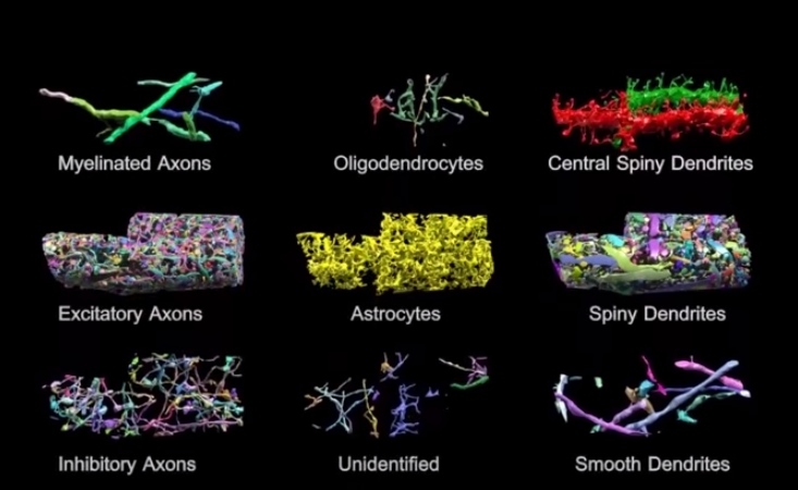
Figure 2: Parts of brain volume (title image) deciphered into various components.
The Human Connectome project at Mass General Hospital (MGH) is working on developing high resolution neuroimaging methods. They’re working on figuring out how varying white matter fiber connectivity correlates to brain function and how cytoarchitectonics (the pattern of neuron organization/density in different areas of the brain, used to decipher Brodmann’s areas in the brain) can influence brain connectivity [2]. Methods they’re using include diffusion spectrum imaging (DSI), which is similar to diffusion tensor imaging (DTI) as both allow scientists to observe white matter connectivity since water diffuses parallel to white matter tracts. DTI measures the preferred and mean direction of the diffusion of water while DSI measures it in many different directions [3]. Scientists at MGH are also working on developing tools for high angular diffusion imaging (HARDI), which “address[es] the challenge of imaging crossing fibers by applying a stronger magnetic gradient for a longer time and capturing many more images of diffusion in various directions” [3].
Researchers are hoping to find patterns of connectivity by deciphering how our 100 trillion neurons are linked. This information could help scientists better understand the roots of different neurological or neurodegenerative diseases where perhaps neural connectivity differs significantly. This is a challenging project but the results of it could be a major breakthrough for neuroscience.
~ Srijesa K.
Sources:
[1] “Untangling the Connectome”
[3] “White’s the Matter: A basic guide to white matter imaging using diffusion MRI”
An Understanding of Language-Processing with Audiobooks
When I read a book such as the one about the romantic life of Peeta and Katniss, I delve into the lives of the characters so much that I worry about myself because I relate myself to the characters' fantastical lives. All readers face this and that is incredible. Neuroscientists like Roel Willems and Annabel Nijhof would completely agree. In fact, they recently published a study revealing the neurological effects of listening to audiobooks.
In the experiment, researchers had the subjects listen to chapters of several different audiobooks and recorded their neurological responses using functional Magnetic Resonance Imaging (fMRI). According to the results, the subjects focused mostly on either the actions of characters or the feelings and intentions of the characters. In the subjects that reported to prefer empathizing with the characters more, the fMRIs showed heightened activity in the anterior medial prefrontal cortex, whereas those that reported enjoying the action aspect of the story more had elevated activity in the motor cortex. Interestingly enough, the subjects that showed higher activation of the anterior medial prefrontal cortex displayed lower activation of the motor cortex when listening to the action parts of the stories. On the contrary, the subjects that engaged more with the actions of the story were applicable. This study provides neurological evidence that people relatively have different strengths and preferences when it comes to reading.
These findings imply this study could be used to know how people engage with literature. If we master what literature people prefer, we might be able to turn literature into a very fun activity. This research also offers further understanding on how we process language. As stated above, it shows that when listening to narration some people focus more on empathizing aspects while others focus more on the action-packed plot. Not only this teaches how we read books, but also how we process written and oral stories.
Source:
The Mentioned Experiment: http://neurosciencenews.com/literature-neuroimaging-psychology-1748/
Is iPhone Separation Anxiety Real?
 Have you ever searched for a ringing phone, feeling your anxiety increase with each ring? Or experienced a mini heart attack when you thought you lost your phone only to discover it was in a different pocket? Most older generations would criticize you for being so obsessed with technology, but recent studies have shown that ‘iPhone separation anxiety’ is a real disorder – and it is plaguing the younger generations of today’s society.
The average person spends about two hours and fifty-seven minutes on a smartphone or tablet every day. Most of us get stressed out if we misplace our phone, are constantly checking for notifications, and even feel ‘phantom vibrations’ – a sensation that we have received a notification when we really have not. Most people would dismiss this anxiety as an unhealthy obsession with technology, but research has shown that these feelings are legitimate and smartphone separation can have serious psychological and physiological effects.
Optogenetics: Using Light to Turn Memories On and Off

Scientists have known about rhodopsins that are responsible for sensing light for a while. What if there was a way to insert those rhodopsins inside neurons? That’s exactly what scientists were experimenting with in the early 2000s and it’s this idea that lead to the birth of optogenetics. By taking the DNA of channel rhodopsins from algae and inserting them into the membrane of neurons, scientists were able to make neurons sensitive to particular wavelengths of light. Channel rhodopsin and halorhodopsin are among the opins inserted into neurons by injecting viruses. Channel rhodopsin activates neurons while halorhodopsin silences them. Once the neuron expresses the light-gated cation channel channelrhodopsin-2 in its cells membrane, shining light on it for as little as a few milliseconds has a profound effect. It causes the opening of the channelrohodopsin-2 molecules, allowing positively charged ions to enter cell and cause the cell to fire.

Check out this video to see how optogenetics works.
Many experiments today use optogenetics to selectively turn neurons on and off in mice. What makes this method mind blowing is the high spatial and temporal resolution it gives scientists when working with the brain. It can be used on neurons in on a petri dish or within a living animal. It could be used to learn more about the function of particular brain regions. For instance, one could temporarily inactivate one region to observe how it impacts activity in other connected brain regions.
Furthermore, it’s minimally invasive: once the virus containing the rhodopsin has been injected, all the scientist needs to do is shine a pulse of light. Researchers at Stanford have used optogenetics to induce muscle contractions in mice. At Case Western Reserve University, researchers implemented it to restore motor function in rats paralyzed by spinal cord injuries. Could optogenetics be used to recover vision loss, something most humans deal with as they age? Experiments conducted on mice with a lack of photoreceptors shows that shining light on bipolar cells (containing channelrhodopsin-2) causes action potentials to fire in the visual cortex. It would be amazing if scientists could overcome biomedical and technical obstacles to make this work in humans too.
Across the river at MIT, members of the Tonegawa lab have taken the technique of optogenetics one step further. Steve Ramirez and Xu Liu have been working to localize memories in the brain and activate them with a light “switch”. And they have accomplished this feat in mice. Promising experiments with mice suggest optogenetics can be used to turn off traumatizing memories and activate pleasant once. This could have implications for PTSD, where horrific memories could be suppressed. They also have experimented with the idea of implanting false memories into the brain, which they call “Project Inception.” For more information on this work (and a good laugh), check out Steve Ramirez and Xu Liu’s TED talk.
Seems to me like optogenetics is a promising technique that can lead to breakthroughs in neuroscience.
-Srijesa K.
Sources
[1] “The Birth of Optogenetics” http://www.the-scientist.com/?articles.view/articleNo/30756/title/The-Birth-of-Optogenetics/
[2] “Potential Benefits of Optogenetics” http://optogenetics.weebly.com/what-is-it1.html
New Huntington’s disease clinical trial to enter Phase 1 in 2015
Huntington’s disease (HD) is a neurodegenerative disorder that slowly diminishes one’s ability to walk, talk, and reason until eventually control is completely lost. HD is an autosomal dominant genetic disorder, so everyone who carries the gene is guaranteed to develop the disease and will have a 50% chance of passing it on. Currently, there is no cure for this devastating disease, and most die within 10-20 years after onset.
With no treatment or way to stop the progression of HD, the outlook of those diagnosed has seemed pretty bleak. Until now. A new clinical trial for an HD drug is set to enter Phase 1 in 2015. This drug is meant to target the source of HD itself: the Huntington gene.
The Miracle of Neurogenesis
Neurogenesis occurs in two areas in the human adult: in the dentate gyrus of the hippocampus and in the olfactory system. The hippocampus is vital to learning new information and memory consolidation, thus it makes sense that new neurons need to be born in that region. The olfactory system is needs neurogenesis to process to new information. Majority of neurogenesis actually occurs during prenatal development. In fact humans initially have more neurons that necessary for survival. Apoptosis (programmed cell death) occurs to prune the synapses established during early development.
Many studies have been conducted that investigate ways to increase neurogenesis. Such activities include voluntary physical exercise or being in enriched environments. Experiments with rats have shown that being in an enriched environment where rats are exposed to complex objects, toys, running wheels, etc. spark improvements in performance of tasks measuring levels of learning and memory. For humans, I suppose an enriched environment could be a place involving novel stimuli or
If you think about it, how could neurogenesis be bad? I mean people with neurodegenerative diseases suffer from the consequences of neuronal loss, right? However, according to a study published in May 2014 in Science, exercise could induce amnesia. An article regarding this study states, “Adult mice that exercised on a running wheel after experiencing an event were more likely than their inactive mates to forget the experience.” Thus it appears that the neurogenesis that occurs during exercise may be “wiping out” neurons that encoded previous memories. Furthermore, when neurogenesis was pharmacologically inhibited scientists observed a recall failure in the rats. This article relates this phenomenon to the fact that children cannot form long term memories until they are 3-4 years of age.
Despite controversy about neurogenesis, olfactory ensheathing cells (OECs) have been used to help a paralyzed man walk once again. An article published in October in BBC News describes how doctors in Poland accomplished this feat. The first step to this process was to extract cells from the patient’s olfactory bulbs (they removed one olfactory bulb and grew the cells in culture). Two weeks later the cells were implanted in the areas surrounding the spinal cord damage the patient had experienced. This action allowed the spinal cord cells to regenerate because the nerve grafts acted “as a bridge to cross the severed cord.” The implications of this type of surgery are pretty amazing – it could work wonders for paralyzed veterans/other individuals and people dealing with dysfunctions relevant to spinal cord damage.
Sources:
Exercise Can Erase Memories - The Scientist
Paralyzed Man Walks Again After Cell Transplant - BBC
- Srijesa K.
Food Addiction- Why You Can’t Eat Just One Oreo

Nothing is better than satisfying a craving for junk food. Be it Oreos, chips, or chocolate, digging into your favorite snack is probably the best feeling in the world. But what happens when junk food stops becoming a guilty pleasure and instead becomes a real addiction?
Research has discovered that sugar is more addictive than cocaine and that your brain becomes addicted to its own release of opioids in the reward system. The reward system in our brains evolutionarily benefited us by rewarding us for engaging in behavior that encouraged our survival. Thus, when we eat, the neurotransmitter dopamine (usually associated with happiness and pleasure) is released, causing the happy feeling you get when you satisfy a craving.
The world that we live in is one of easy access to sugar-rich foods and has lead to the normalization of overly sweet and processed foods in our everyday lives. Just like any addiction, tolerance to a substance may occur. In this case, constantly activating the reward system and releasing dopamine by continuously eating junk food can cause dopamine receptors to down-regulate. At this point, dopamine receptors are removed by the brain to maintain a balanced state. Fewer dopamine receptors prevent the same stimulation/effect created by dopamine release; thus we tend to eat more junk food to reach the same level of pleasure as before.
More
Is the brain the only place that stores our memories?
 Do you ever think about your childhood or replay an event in your head that happened 15 years ago but its so vivid that it seems like it happened yesterday? Do you ever hear something and think it sounds like your favorite song and then start singing that song? These are memories that were formed in your brain that are replayed as a result of a specific stimulus. For a long time scientists believed that memories were formed, processed, and sent to different destinations in the brain. Dr. Wilder Penfield was one of the first to accidentally discover this. In the 40s he electrically stimulated different areas of his patients' brains while they were under local anesthesia and found that the region he stimulated would elicit specific memories in the patient's life (see video below). For example, in one of his patients he stimulated her temporal lobe (auditory cortex) and she started to hum her favorite song out loud. This suggested that the memory of this song was stored in the place where it was processed or originated (i.e. the auditory cortex processed the first time she listened to the song). Penfield concluded that the cortex (the outer layers of the brain) stored the "complete record of the stream of consciousness; all those things in which a man was aware at any time..." Until recently, scientists have believed this phenomenon.
An Introduction to Sensory Processing Disorder

Sensory Processing Disorder(SPD) is a disorder that impairs processing of sensory information in the brain. For children it can cause klutziness, an inability to properly orient the body, poor fine motor skills, and a hypersensitivity to sound. In summary, this disorder makes it very difficult to process incoming information. The condition is very common for children and often is not properly diagnosed because until recently, there had not been a real biological explanation for the disorder. A very common treatment for these patients is occupational therapy.
The Science Behind the Fear

We all feel scared, jumpy, or anxious but have you ever stopped to think why? Have you ever wondered why you get those goosebumps or why you feel that you are frozen sometimes? Just in time for Halloween, here is some information on how our body processes fear. According to NIMH, fear is defined as a feeling of disquiet that begins rapidly in the presence of danger and dissipates quickly once the threat is removed and is also generally adaptive. Fear is a primitive response.
While neuroscientists do not understand the pathways that we take to interpret fear, there are a few recent studies that examine this. In the first one, scientists identified specific neurons called SOM+ that are linked to a type of “fear memory” held in the amygdala.
