Our Research
We use brain imaging, non-invasive brain stimulation, and behavioral assessments to understand how early experiences shape motor development.

The DEMR Lab uses a variety of techniques including brain imaging, non-invasive brain stimulation, and behavioral assessments to understand motor development and brain function. In particular, we are interested in studying how sensorimotor experiences shape the development of motor circuits involved in the control of the hands and arms.
Our goal is to use this information to develop targeted therapies for children with cerebral palsy.
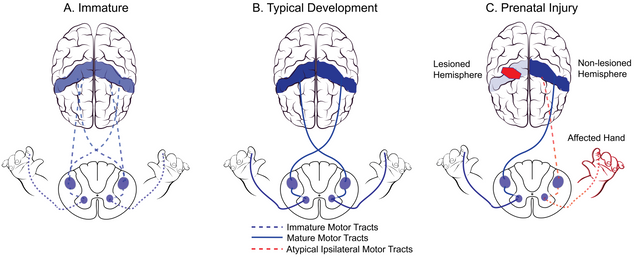
Model of Corticospinal Tract (primary pathway for skilled movement) Development
Brain Imaging
Functional Near-Infrared Spectroscopy (fNIRS)
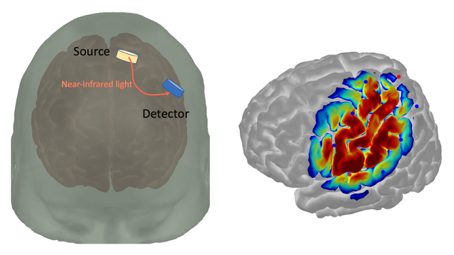
How fNIRS works
fNIRS is a non-invasive method for studying functional brain activity. Given the relatively easy set-up of this imaging tool — a cap worn on the head — fNIRS is particularly suited for measuring brain function in infants and children.
We use fNIRS to study: (1) how spontaneous infant movements shape brain activity, and (2) how movement therapy improves brain function in older children. We work closely with collaborators at the BU Neurophotonics Center to deploy state-of-the-art brain imaging technologies.
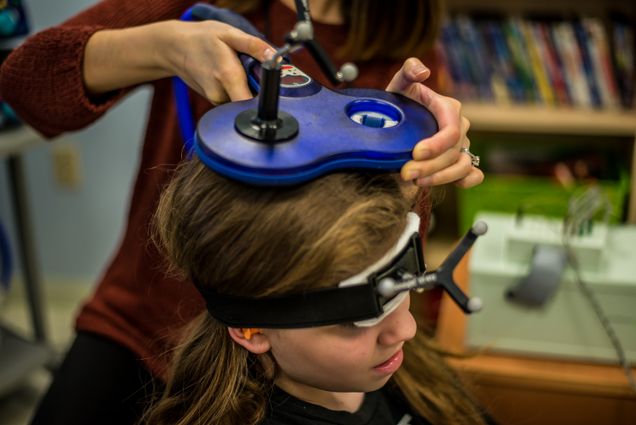
For families: The fNIRS cap looks like a helmet with small sensors. It is completely safe and painless — your child simply wears the cap while playing with toys or moving naturally.


Brain Stimulation
Transcranial Magnetic Stimulation (TMS)
TMS is a form of non-invasive brain stimulation that can be used to map brain areas responsible for the control of the hands. We use TMS to probe the integrity and excitability of motor systems following intensive rehabilitation.
In our lab, TMS is done at rest: children can relax and watch a movie while we perform the measurements.
For families: TMS uses a small magnetic coil held near the head. It produces a brief clicking sound and a gentle tapping sensation. Children find it easy to tolerate, and sessions are kept short and comfortable.
We encourage you to watch a video made by our collaborator Dr. Kathleen Friel featuring children describing what it feels like to be part of a TMS study.

Brain Imaging
Diffusion Tensor Imaging (DTI)
DTI is a brain imaging technique that enables the measurement of white matter pathways. We use DTI to understand how brain lesions that occur early in development affect the organization of motor and sensory pathways.
By understanding how these pathways reorganize, we hope to discover mechanisms of plasticity that can support adaptive motor function following developmental brain injury.
For families: DTI is performed inside an MRI scanner. It is safe and painless — no injections or radiation are involved. The scan takes place while your child lies still while watching a movie, and we make the experience as comfortable as possible.
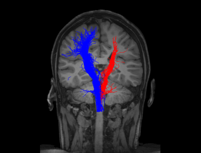
DTI of motor pathways (blue = unaffected, red = affected)

Our child-friendly mock MRI scanner — kids can practice before the real scan
Collaborators
Our research involves collaborations with other researchers and centers: