Publications
Selected journal publications:
Kim, B.K., Howard, A.C., Cheng, T.Y. et al. Mapping human cerebral blood flow with high-density, multi-channel speckle contrast optical spectroscopy. Commun Biol 8, 1553 (2025). https://doi.org/10.1038/s42003-025-08915-x
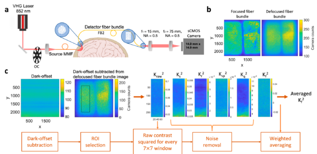
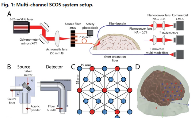 Recently, speckle contrast optical spectroscopy (SCOS) enabled non-invasive, high signal-to-noise-ratio (SNR) human cerebral blood flow (CBF) measurements, relevant for both neuroscience and clinical monitoring of diseases with CBF dysregulation. Single-channel SCOS measurements limit the information obtained to only one location on the head. In this work, we develop a multi-channel SCOS system to map spatial heterogeneity in CBF changes during human brain activation. Using a galvanometer, we temporally multiplex a free-space laser to 7 source fibers positioned at different locations on the head. Diffuse light collected from the tissue is captured by fiber bundles projecting to 17 complementary metal-oxide semiconductor (CMOS) cameras, resulting in 50 source-detector channels measuring optical density (OD) and relative CBF changes covering an area of 7.6 cm by 6.6 cm on the head. We validate the spatial specificity and stability of the system using a liquid flow phantom. We then measure brain activity during a word-color Stroop task in 15 subjects and obtain brain activation maps. The average signal changes in the channel showing the largest activation is 0.017 in ΔOD and 6.6% in CBF
Liu, B., Tang, R.P., Long, E.A., Yaqoob, Z., Simkulet, M.G., Postnov, D.D., Erdener, Ş.E., Boas, D.A. and Cheng, X., 2025. Mapping of blood flow and slow speckle tissue dynamics using laser speckle contrast imaging. Biomedical Optics Express, 16(9), pp.3712-3724.
 Laser speckle contrast imaging (LSCI) is a wide-field optical technique commonly used to monitor cerebral blood flow (CBF). Recently, we have discovered that besides the fast-decorrelating signals from blood flow, LSCI can also detect slow-decorrelating signals associated with cellular activity. The ability to image these signals has significant implications for various research areas such as ischemic stroke, as it enables longitudinal monitoring of both vascular and cellular dynamics, offering new biomarkers for tissue viability, injury progression, and therapeutic response. Here, we demonstrated that epi-illumination LSCI enables the mapping of both slow speckle dynamics (SSD) for evaluating cellular dynamics and traditional fast speckle dynamics (FSD) for evaluating CBF. We found that SSD signals are much more evident with epi-illumination than with conventional oblique illumination LSCI. Using mouse models of ischemic stroke, including both permanent and transient occlusion of the distal middle cerebral artery (dMCA), we demonstrated the system’s ability to track stroke progression from minutes to days post-stroke. This study establishes a powerful, label-free imaging tool for investigating both cellular and vascular health during stroke core evolution.
Howard, Alexander C., Byungchan Kim, Laura Carlton, Meryem A. Yücel, Bingxue Liu, David A. Boas, and Xiaojun Cheng. “Validation of the Linearity in Image Reconstruction Methods for Speckle Contrast Optical Tomography.” IEEE Journal of Selected Topics in Quantum Electronics (2025).
This work demonstrates that linear methods, such as the general linear models developed for fNIRS, can be applied for SCOS/SCOT data processing.
Speckle contrast optical spectroscopy (SCOS) is an optical technique capable of measuring human cerebral blood flow and brain function non-invasively. Its tomographic extension, speckle contrast optical tomography (SCOT), can provide blood flow variation maps with measurements using overlapping source-detector channel pairs. Linearity is often assumed in most image reconstruction methods, but non-linearity could exist in the relations between measured signals and blood flow variations. We have constructed a forward model for SCOT using the Rytov approximation to solve the correlation diffusion equation and compared it with the first Born approximation as well as the more accurate, but computationally expensive Monte Carlo simulation approach. We have shown that the results obtained using the Rytov approximation are in good agreement with the Monte Carlo simulations, while the first Born approximation deviates from the other two methods for large blood flow variations. For instance, the first Born approximation breaks down at around 30% cerebral blood flow (CBF) changes within a volume of size , therefore we recommend using the Rytov approximation above this threshold. We have shown that our defined blood flow index (BFi) measured in SCOT is linearly related to local CBF variations, thus the forward and inverse problems can be solved linearly using the sensitivity matrix approach. We have then demonstrated image reconstruction experimentally showing human brain activations using our recently developed high-density SCOS system. Our method guides experimental system design and data analysis for SCOT.
Cheng, Tom Y., Byungchan Kim, Bernhard B. Zimmermann, Mitchell B. Robinson, Marco Renna, Stefan A. Carp, Maria Angela Franceschini, David A. Boas, and Xiaojun Cheng. “Choosing a camera and optimizing system parameters for speckle contrast optical spectroscopy.” Scientific Reports 14, no. 1 (2024): 11915.
 Speckle contrast optical spectroscopy (SCOS) is an emerging camera-based technique that can measure human cerebral blood flow (CBF) with high signal-to-noise ratio (SNR). At low photon flux levels typically encountered in human CBF measurements, camera noise and nonidealities could significantly impact SCOS measurement SNR and accuracy. Thus, a guide for characterizing, selecting, and optimizing a camera for SCOS measurements is crucial for the development of next-generation optical devices for monitoring human CBF and brain function. Here, we provide such a guide and illustrate it by evaluating three commercially available complementary metal–oxide–semiconductor cameras, considering a variety of factors including linearity, read noise, and quantization distortion. We show that some cameras that are well-suited for general intensity imaging could be challenged in accurately quantifying spatial contrast for SCOS. We then determine the optimal operating parameters for the preferred camera among the three and demonstrate measurement of human CBF with this selected low-cost camera. This work establishes a guideline for characterizing and selecting cameras as well as for determining optimal parameters for SCOS systems.
Bingxue Liu, Dmitry Postnov, David A. Boas, and Xiaojun Cheng, 2024. Dynamic light scattering and laser speckle contrast imaging of the brain: theory of the spatial and temporal statistics of speckle pattern evolution. Biomed. Opt. Express 15(2), 579-593.
Dynamic light scattering (DLS) and laser speckle contrast imaging (LSCI) are closely related techniques that exploit the statistics of speckle patterns which can be utilized to measure cerebral blood flow (CBF). Conventionally, the temporal speckle intensity auto-correlation function is calculated in DLS, while the spatial speckle contrast is calculated in LSCI measurements. Due to the rapid development of CMOS detection technology with increased camera frame rates while still maintaining a large number of pixels, the ensemble or spatial average of the auto-correlation functions as well as the temporal contrast can be easily calculated and utilized to quantify CBF. Although many models have been established, a proper summary is still lacking to fully characterize DLS and LSCI measurements for spatial and temporal statistics, laser coherence properties, various motion types, etc. As a result, there are many instances where theoretical models are misused. Therefore, we aim to provide a review of the speckle theory for both DLS and LSCI measurements with detailed derivations from first principles, taking into account non-ergodicity, spatial and temporal statistics of speckles, scatterer motion types, and laser coherence properties. From these calculations, we elaborate on the differences between spatial and temporal averaging for DLS and LSCI measurements that are typically ignored but can result in inaccurate measurements of blood flow. We also obtained in vivo mouse brain measurements using high frame rate CMOS cameras which have not been demonstrated before. This work provides a useful guide for choosing the correct model to analyze spatial and temporal speckle statistics in in vivo DLS and LSCI measurements.
Kim, B., Zilpelwar, S., Sie, E.J., Marsili, F., Zimmermann, B., Boas, D.A. and Cheng, X., 2023. Measuring human cerebral blood flow and brain function with fiber-based speckle contrast optical spectroscopy system. Communications Biology, 6, 844.
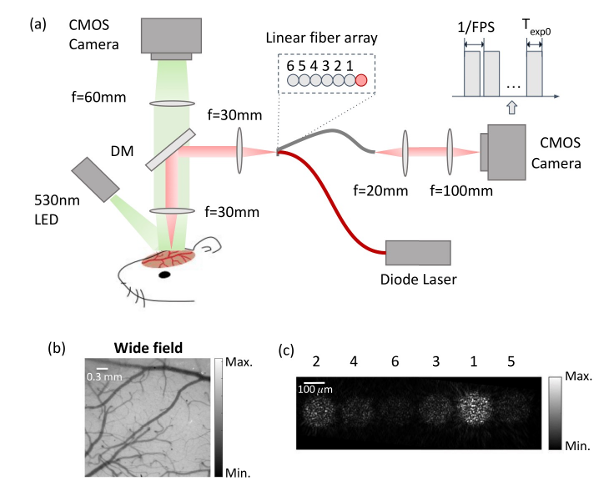
Cerebral blood flow (CBF) is crucial for brain health. Speckle contrast optical spectroscopy (SCOS) is a technique that has been recently developed to measure CBF, but the use of SCOS to measure human brain function at large source-detector separations with comparable or greater sensitivity to cerebral rather than extracerebral blood flow has not been demonstrated. We describe a fiber-based SCOS system capable of measuring human brain activation-induced CBF changes at 33 mm source-detector separations using CMOS detectors. The system implements a pulsing strategy to improve the photon flux and uses a data processing pipeline to improve measurement accuracy. We show that SCOS outperforms the current leading optical modality for measuring CBF, i.e. diffuse correlation spectroscopy (DCS), achieving more than 10x SNR improvement at a similar financial cost. Fiber-based SCOS provides an alternative approach to functional neuroimaging for cognitive neuroscience and health science applications.
Liu, B., Shah, S., Küreli, G., Devor, A., Boas, D.A. and Cheng, X., 2023. Measurements of slow tissue dynamics with short-separation speckle contrast optical spectroscopy. Biomedical Optics Express, 14(9), pp.4790-4799.
Laser speckle contrast imaging (LSCI) measures 2D maps of cerebral blood flow (CBF) in small animal brains such as mice.
The contrast measured in LSCI also includes the static and slow-varying components that contain information about brain tissue dynamics. But these components are less studied as compared to the fast dynamics of CBF. In traditional wide-field LSCI, the contrast measured in the tissue is largely contaminated by neighboring blood vessels, which reduces the sensitivity to these static and slow components. Our goal is to enhance the sensitivity of the contrast to static and slow tissue dynamics and test models to quantify the characteristics of these components. To achieve this, we have developed a short-separation speckle contrast optical spectroscopy (ss-SCOS) system by implementing point illumination and point detection using multi-mode fiber arrays to enhance the static and slow components in speckle contrast measurements as compared to traditional wide-field LSCI (WF-LSCI). We observed larger fractions of the static and slow components when measured in the tissue using ss-SCOS than in traditional LSCI for the same animal and region of interest. We have also established models to obtain the fractions of the static and slow components and quantify the decorrelation time constants of the intensity auto-correlation function for both fast blood flow and slower tissue dynamics.
Using ss-SCOS, we demonstrate the variations of fast and slow brain dynamics in animals before and post-stroke, as well as within an hour post-euthanasia. This technique establishes the foundation to measure brain tissue dynamics other than CBF such as intracellular motility.
Zilpelwar, S., Sie, E.J., Postnov, D., Chen, A.I., Zimmermann, B., Marsili, F., Boas, D.A. and Cheng, X., 2022. Model of dynamic speckle evolution for evaluating laser speckle contrast measurements of tissue dynamics. Biomedical Optics Express, 13(12), pp.6533-6549.
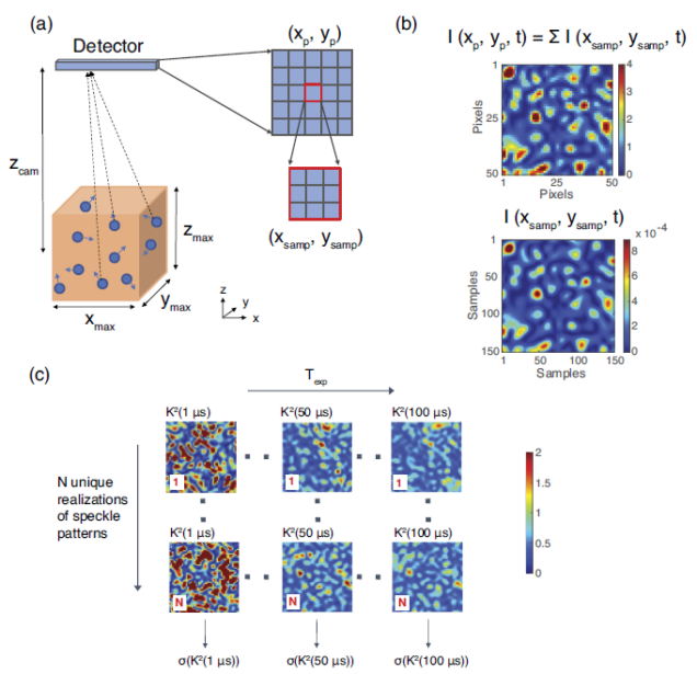
We introduce a dynamic speckle model (DSM) to simulate the temporal evolution of fully developed speckle patterns arising from the interference of scattered light reemitted from dynamic tissue. Using this numerical tool, the performance of laser speckle contrast imaging (LSCI) or speckle contrast optical spectroscopy (SCOS) systems which quantify tissue dynamics using the spatial contrast of the speckle patterns with a certain camera exposure time is evaluated. We have investigated noise sources arising from the fundamental speckle statistics due to the finite sampling of the speckle patterns as well as those induced by experimental measurement conditions including shot noise, camera dark, and read noise, and calibrated the parameters of an analytical noise model initially developed in the fundamental or shot noise regime that quantifies the performance of SCOS systems using the number of independent observables (NIO). Our analysis is particularly focused on the low photon flux regime relevant for human brain measurements, where the impact of shot noise and camera read noise can become significant. Our numerical model is also validated experimentally using a novel fiber-based SCOS (fb-SCOS) system for a dynamic sample. We have found that the signal-to-noise ratio (SNR) of fb-SCOS measurements plateaus at a camera exposure time which marks the regime where shot and fundamental noise dominate over camera read noise. For a fixed total measurement time, there exists an optimized camera exposure time if temporal averaging is utilized to improve SNR. For a certain camera exposure time, photon flux value, and camera noise properties, there exists an optimized speckle-to-pixel size ratio (s/p) at which SNR is maximized. Our work provides the design principles for any LSCI or SCOS systems given the detected photon flux and properties of the instruments, which will guide the way for the experimental development of a high-quality, low-cost fb-SCOS system that monitors human brain blood flow and functions in the future.