Imaging through a single optical fiber

Microscope techniques to image inside tissue are generally limited by poor depth penetration. Microendoscopy, wherein a probe is physically inserted into the tissue, can overcome this limitation in depth penetration, but at the expense of invasiveness and tissue damage due to the size of the probe. Our goal here is to palliate these problems by developing an ultra-miniature microendoscope probe based on a single, lensless optical fiber.

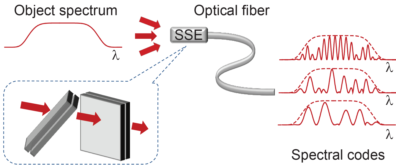
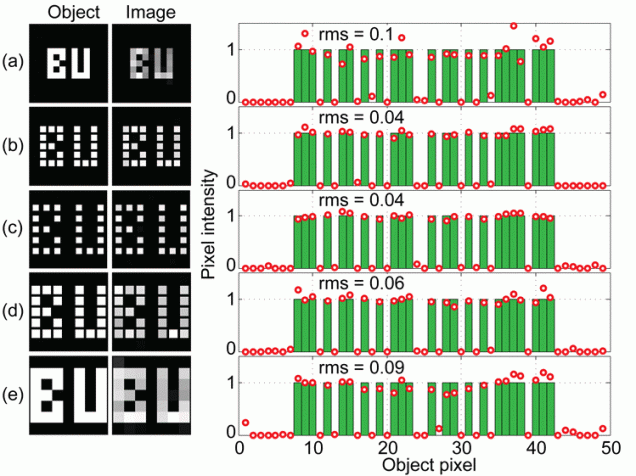
The direct transmission of an image through an optical fiber is difficult because spatial information becomes scrambled upon propagation. We have recently demonstrated an image transmission strategy where spatial information is first converted to spectral information. Our strategy is based on a principle of spread-spectrum encoding, borrowed from wireless communications, wherein object pixels are converted into distinct spectral codes that span the full bandwidth of the object spectrum. Image recovery is performed by numerical inversion of the detected spectrum at the fiber output. We have provided a simple demonstration of spread-spectrum encoding using macroscopic Fabry-Perot etalons. Our technique enables the 2D imaging of luminous (i.e. incoherent) objects with high throughput independent of pixel number. Moreover, it is insensitive to fiber bending, contains no moving parts, and opens the attractive possibility of extreme miniaturization down to the size of a single optical fiber.
- R. Barankov, J. Mertz, “High throughput imaging of self-luminous objects through a single fibre”, Nat. Commun. 5:5581 doi: 10.1038/ncomms6581 (2014). link