Laser speckle contrast imaging
Summary: Laser speckle contrast imaging (LSCI) can be used to evaluate blood flow based on spatial or temporal speckle statistics. We describe novel strategies for evaluating the speckle correlation time that are insensitive to detection noise (spatial covariance) and do not require numerical fitting of data (direct characterization). In addition, we describe a technique to correct for bias errors when evaluating speckle correlation time that result from limited sampling statistics. The accuracy of the correlation time is undermined by out-of-focus image blur. We show how the fraction of dynamic versus static light scattering is dependent on focus, and describe a deconvolution strategy to correct for out-of-focus blur. With the aid of a z-splitter prism, which enables instantaneous multifocus imaging, we demonstrate depth-resolved LSCI that can robustly extract multi-plane structural and flow-speed information simultaneously. These methods are applied to in vivo imaging of blood vessels in a mouse cortex and provide improved estimates of blood flow speed.
- S. Zheng, I. Davison, A. Garrett, X. Lin, N. Chitkushev, D. Roblyer, J. Mertz, “Robust speckle contrast imaging based on spatial covariance”, Optica 11, 1733-1741 (2024)
- S. Zheng, J. Mertz, “Correcting sampling bias in speckle contrast imaging”, Opt Lett. 47, 6333-6336 (2022). link
- S. Zheng, J. Mertz, “Direct characterization of tissue dynamics with laser speckle contrast imaging”, Biomed. Opt. Exp. 13, 4118-4133 (2022). link
- S. Zheng, S. Xiao, L. Kretsge, A. Cruz-Martin, J. Mertz, “Depth resolution in multifocus laser speckle contrast imaging”, Opt. Lett. 46, 5059-5062 (2021). link
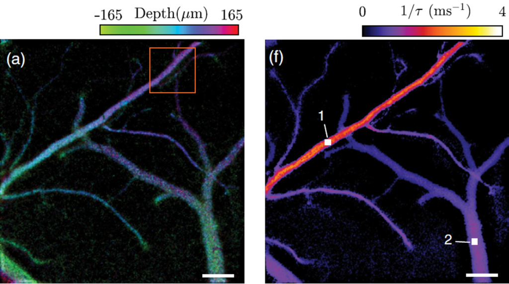
Left: Dynamic fraction associated with blood vessels; Right: Inverse speckle correlation time