Extended-depth-of-field microscopy

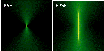
Description: We present a wide-field fluorescence microscopy add-on that provides a fast, light-efficient extended depth-of-field (EDOF) using a deformable mirror with an update rate of 20 kHz. Out-of-focus contributions in the raw EDOF images are suppressed with a deconvolution algorithm derived directly from the microscope 3D optical transfer function. Demonstrations of the benefits of EDOF microscopy are shown with GCaMP-labeled mouse brain tissue.
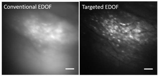
Significantly increased contrast can be obtained by combining EDOF imaging with targeted illumination. The latter strategy involves delivering illumination only to in-focus structure during the focal sweep, using illumination masks controlled by a fast DMD.

- S. Xiao, H. Tseng, H. Gritton, X. Han, J. Mertz, “Video-rate volumetric neuronal imaging using 3D targeted illumination”, Sci. Reports 8, 7921 (2018). link
- W. J. Shain, N. A. Vickers, J. Li, X. Han, T. Bifano, J. Mertz, “Axial localization with modulated-illumination extended-depth-of-field microscopy”, Biomed. Opt. Express 9, 1771-1782 (2018). link
- W. J. Shain, N. A. Vickers, A. Negash, T. Bifano, A. Sentenac, J. Mertz, “Dual fluorescence-absorption deconvolution applied to extended-depth-of-field microscopy”, Opt. Lett. 42, 4183-4186 (2017). link
- W. J. Shain, N. A. Vickers, B. G. Goldberg, T. Bifano, J. Mertz, “Extended depth of field microscopy with a high-speed deformable mirror”, Opt. Lett. 42, 995-998 (2017). link