Dynamic speckle illumination microscopy

Dynamic speckle illumination (DSI) microscopy provides fluorescence sectioning with illumination patterns that are neither well defined nor controlled. The idea of this technique is to illuminate a fluorescent sample with random speckle patterns obtained from a laser. Speckle patterns are granular intensity patterns that exhibit inherently high contrast. Fluorescence images obtained with speckle illumination are therefore also granular; however, the contrast of the observed granularity provides a measure of how in focus the sample is: high observed contrast indicates that the sample is dominantly in focus, whereas low observed contrast indicates it is dominantly out of focus. The observed speckle contrast thus serves as a weighting function indicating the in-focus to out-of-focus ratio in a fluorescence image.
A key feature of speckle illumination is that while the exact intensity pattern incident on a sample is not known, the statistics of the intensity distribution are well known to obey a negative exponential probability distribution (provided the speckle is fully developed). According to this distribution, the contrast of a speckle pattern scales with average illumination intensity. Thus, weighting a fluorescence image by the observed speckle contrast is equivalent to weighting it by the average illumination intensity (as in standard imaging), however with the benefit that the weighting preferentially extracts only in-focus signal.

A second key feature of speckle illumination is that its statistics are invariant even in a scattering medium, since unpredictable phase shifts induced by the medium only further randomize an already randomized laser phase front. Hence fluorescence sectioning based on speckle illumination is robust, since it is insensitive to scattering, aberrations, etc., in the illumination path.
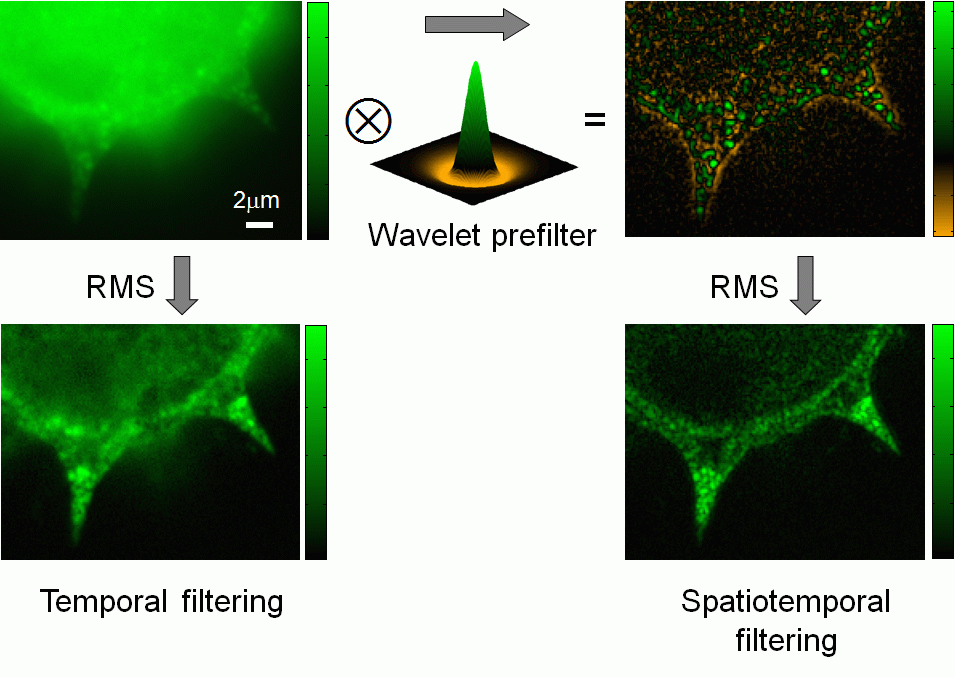
- C. Ventalon, R. Heintzmann, J. Mertz, “Dynamic speckle illumination microscopy with wavelet prefiltering”, Opt. Lett. 32, 1417-1419 (2007). link
- C. Ventalon, J. Mertz, “Dynamic speckle illumination microscopy with translated versus randomized speckle patterns “, Opt. Express 14, 7198-7209 (2006). link
- C. Ventalon, J. Mertz, “Quasi-confocal fluorescence sectioning with dynamic speckle illumination“, Opt. Lett. 30, 3350-3352 (2005). link