Autoconfocal microscopy

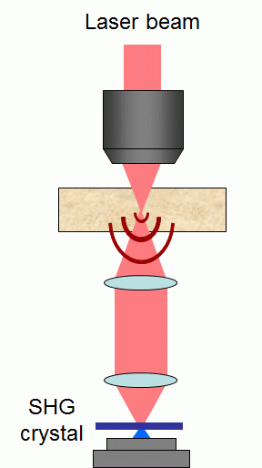
ACM is a nonlinear-optical microscopy that uses a femtosecond-pulsed infrared laser beam to trans-illuminate a tissue sample. As opposed to other nonlinear-optical techniques such as two-photon excited fluorescence (TPEF) microscopy, the laser beam does not generate signal within the sample. Instead, the laser beam traverses the sample and is then re-focused onto a nonlinear crystal. The detected signal is the SHG produced by the crystal. Because this signal is nonlinear, the crystal acts as a “virtual pinhole” and an ACM exhibits 3D resolution and background rejection similar to a transmission-confocal microscope. An ACM requires no sample labeling, is technically simple, and can be readily combined with existing nonlinear imaging modalities such as TPEF microscopy.
We have shown that the equivalent effect of a “virtual pinhole” can be achieved by thermionic emission in a standard PMT photocathode. Because of the nonlinear dependence of thermionic emission on absorbed laser power, a PMT is found to produce a virtual pinhole effect that rejects unfocused light at least as strongly as a physical pinhole. This virtual pinhole effect can be exploited in a scanning transmission confocal microscope equipped with a cw laser source. Because the area of the PMT photocathode is large, signal de-scanning is not required and thermionic detection acts as a self-aligned pinhole.

- D. Lim, K. K. Chu, J. Mertz, “Autoconfocal microscopy with a continuous-wave laser and thermionic detection”, Opt. Lett. 33, 1345-1347 (2008). link
- K. K. Chu, R. Yi, J. Mertz, “Graded-field autoconfocal microscopy”, Opt. Express 15, 2476-2489 (2007). link
- T. Pons, J. Mertz, “Autoconfocal microscopy using nonlinear transmitted light detection”, J. Opt. Soc. Am. B 21, 1486-1493 (2004). link