By fdevita
Spatial Cognition in Philosophy and Neuroscience
In this post, I attempt to present two major metaphysical accounts of space by Kant and Leibniz, then present some recent findings from cognitive neuroscience about the neural basis of spatial cognition in an attempt to understand more about the nature of space and the possible connection of philosophical theories to empirical observations.
Immanuel Kant’s account of space in his Prolegomena serves as a cornerstone for his thought and comes about in a discussion of the transcendental principles of mathematics that precedes remarks on the possibility of natural science and metaphysics. Kant begins his inquiry concerning the possibility of ‘pure’ mathematics with an appeal to the nature of mathematical knowledge, asserting that it rests upon no empirical basis, and thus is a purely synthetic product of pure reason (§6). He also argues that mathematical knowledge (pure mathematics) has the unique feature of first exhibiting its concepts in a priori intuition which in turn makes judgments in mathematics ‘intuitive’ (§7.281). For Kant, intuition is prior to our sensibility and the activity of reason since the former does not grasp ‘things in themselves,’ but rather only the things that can be perceived by the senses. Thus, what we can perceive is based on the form of our a priori intuition (§9). As such, we are only able to intuit and perceive things in the world within the framework naturally provided by the capabilities and character (literally the under–standing) of our understanding. Kant then takes our intuitions of space (and time) as concepts integral to pure mathematics and as necessary components of our intuition (§10.91). More
Do You See What I See?
Philosophy of Mind came into its most compelling forms during the age of modern philosophy beginning with René Descartes. Perhaps infamously, Descartes claimed that mind and body are two distinct substances – philosophical jargon for what exists without the aid of any other thing. For Descartes, the world was clearly and distinctly physical in one sense and entirely mental in another. This seems perplexing, and Descartes did concede that the mind and body were closely intertwined and appeared to act with respect to one another, but his arguments clearly press that they are not causally connected in any way. These notions of dualism seem nearly preposterous with the advent of modern science, but were nonetheless important in developing our thought about the mind in the modern era.
Dualism gave rise to other interesting, yet now strongly refuted movements. One of these was idealism, or the doctrine argued famously by George Berkeley that states that all that exists are either ‘ideas’ or minds that perceive them. In this sense, an idea is defined as that which is perceived, inclusive of information imprinted on the senses, passions and operations of the mind, and conceptions formed by imagination and memory. Importantly, Berkeley argues that these ideas exist ‘in the mind’ exclusively: that is, they are purely mental and all things are simply combinations and aggregations of ideas. These immaterial ‘ideas’ then, are the only objects of human knowledge under idealism, and this theory denies the existence of physical objects entirely! The notion seems preposterous, but there is a very interesting argument found within idealism that can throw our conception of perception for quite the proverbial loop. More
Genome or Connectome?
Sebastian Seung is a professor of computational neuroscience and physics at MIT. His research in the neuroscience field involves "connectomes," or the map of connections between and among neurons. The endeavor of investigating and mapping connectomes began in the 1980s and jumped off with the elucidation of the complete connectome of the worm C. elegans in 1986. While C. elegans has about 300 neurons, humans have about 10 billion neurons and ten times that number of connections. These connections can grow and change with and from neural activity and experience, combining to permutations exponentially greater that those of DNA and its four bases. Seung proposes that we "are our connectomes" rather than our genomes, implying that our thoughts, experiences, emotions, and consciousness itself may have a purely neural basis. To refrain from any more spoilers, he artfully expands and explains his hypothesis in the above TED talk that it is surely worth viewing. For a greater philosophical inquiry inspired by his ideas, is our matter all that matters?
This Is Your Brain on RF-EMF
Can you hear me now? For years, it has been popular doctrine that cell phone use is bad for our brains, but we glue our phones to our ears anyway. Cell phones emit radio frequency-modulated electromagnetic fields (RF-EMFs) that are questioned for their potential danger when the brain is exposed to them. The oscillatory frequencies of RF-EMFs correspond to those measured in neural tissue, and thus could interfere with neural activity. The amount of electromagnetic radiation given off by our communication devices is small, but is radiation all the same. Radiation exposure is dangerous for any kind of cell in our body, and can penetrate cells and damage DNA either by crashing into the molecule directly or causing damage indirectly by forming free radicals from water that can have cancer-causing effects.

Knocking Out Parkinson's Disease
![]()
Parkinson's disease is one of the most infamous neurological disorders known to medicine and has afflicted many, including Muhammed Ali and Michael J. Fox. Described by it's characteristic tremors and shaking, the disease induces loss of control of motor function as a result of neuronal death in dopamine-releasing (dopaminergic) neurons in a midbrain structure called the substantia nigra. In the pathology of Parkinson's Disease, this localized population of dopaminergic neurons start to die, while other neurons in the substantia nigra and other local dopaminergic neurons remain healthy and functional. This has puzzled researchers extensively, but a recent letter to Nature provides some breakthroughs in understanding what is happening in these degenerative neurons.
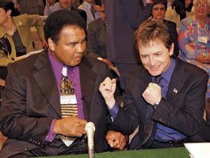
Muhammed Ali and Michael J. Fox are two celebrities battling Parkinson's Disease.
Recent research into Parkinson's suggests that the problems are localized to the mitochondria of the suspect neurons, leading to the conclusion that the disease is related to problems with metabolism. The mitochondria are the machines of cell metabolism and the stage for the Kreb's Cycle and the Electron Transport Chain (ETC), which are the two major energy producing processes of the cell. The metabolism occurring in the mitochondria produces large amounts of adenosine triphosphate (ATP) which is used as energy currency in all cells of the body, and also lead to the production of water to be used elsewhere by the cell. Oxidative stress in the water producing portion of the metabolic pathway can lead to the production of oxygen free radicals, which are highly reactive as a result of a single free electron (free radicals have implications in other degenerative disorders and cancer). Scientists now suspect that these free radicals are contributing to the neurodegenerative consequences of Parkinson's Disease.
Neuroimaging study showing the decreased neuronal activity in Parkinson's Disease.
In the Nature study, scientists tagged the mitochondria of the Parkinson's suspect neurons with fluorescent protein that would allow them to observe oxidation states of the cells in rats. They found that Parkinson's cells were in fact under a high level of oxidative stress and showed stress patterns at regular intervals, concluding that the cells function rhythmically when releasing dopamine. This rhythmic function is associated with ATP driven increases of calcium levels in the neurons, and the study suggests that this rhythmic calcium fluctuation is the instigator of the oxidative stress associated with Parkinson's disease. In further experimentation, the researchers blocked these calcium influxes with drugs, which led to a decrease in Parkinson's-like oxidative stress in mouse models of early-onset Parkinson's disease. These drugs are known to be tolerated by humans well, and provide a legitimate option for medicinal therapy in the disease.
Oxidant stress evoked by pacemaking in dopaminergic neurons is attenuated by DJ-1 - Nature 2010
Mind the Gap
![]()
The discoveries of modern neuroscience have certainly heightened our understanding of the brain and its functions, and have begun to provide us with a physical groundwork for the complicated problem of effectively investigating the mind. While it is certainly beneficial to establish physical principles that underly cognitive function of the brain, how does this effect the larger endeavor of understanding the mind? Neuroscientists such as Rebecca Saxe of MIT are converging on things like the nuroanatomical basis of moral judgment and just scraping the surface of what can bridge the gap between what physically "is" and what metaphysically "ought" to be. In her experiments, Saxe proposes that she has pinpointed the right temporoparietal junction (RTPJ) as a brain center for making moral judgments and has conducted experiments with magnetic brain stimulation that can effectively change the moral judgments of her subjects. Please see her TED talk here for a full explanation of her study.
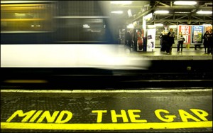
Crossing the gap at the wrong time can be devastating.
In the 1700s, David Hume proposed what has now become known widely as the Is-Ought Problem. He calls for caution in making statements about morality or what "ought" to be based on extrapolations of what "is" and that what ought to be does not necessarily follow from what is. The problem aptly applies to neuroscientists like Saxe whose research make strong suggestions about the neural basis of existence and attempts to bridge the is-ought gap. All of this research is establishing a large library of what "is" concerning the brain, but it also suggests that metaphysical concepts such as morality and meta-ethics can be reduced to neurological connections and connectivity. Hume stresses that while what is and what ought to be are important revelations in and of themselves, what ought to be need not follow from what is. Neuroscience must understand this separation as its advances begin to encroach on many of philosophy's already well-established concepts.
Brain activity is only one component of our consciousness.
What I'm saying here is that modern neuroscience must use caution in making conclusions about human nature. Empirical evidence can certainly be used to help understand more abstract ideas, but the evidence and the ideas must remain seperate with respect to causality. Making discoveries about brain function and the empirical science behind things like emotion or judgment is a valiant and respectable scientific investigation. However, this pursuit must be kept separate and distinct from the pursuit of understanding how we ought to be or act. Our moral thought is something more abstract and multidimensional than connections between neurons and sequential acton potentials. While investigation of the science of the mind is important, it should not seek to explain our existence nor try to answer philosophy's greatest problems with calculations and empirical data.
For reference:
The Is-Ought Problem - David Hume via Wikipedia
Theory of Mind TED Talk - Rebecca Saxe (MIT)
David Hume, Meta-Ethics - Wikipedia
The Creator Inside Our Minds
![]()
Through humanity's existence there has been a backbone of culture, tradition and out of that has come religion. Since ancient times, humans as a species have devoted their time and mental capacity to a higher being that we cannot physically perceive nor interact with. There are varying viewpoints from different disciplines that try to explain the nature of this God and where he or she resides, if anywhere. While many have displaced intelligent design as a plausible theory, mathematics offers that there may be divine math behind our existence, theoretical physics has routinely said that particle dynamics can answer our questions, while biochemistry presents the primordial soup theory and DNA replication. However, there are people among us who claim to have had "mystical experiences" that put them in touch with this ever elusive God on a level experienced by few.
The Big Bang Theory is a commonly accepted scientific theory of creation
Now, neuroscience has something to say about this "God experience." Michael Persinger, a cognitive scientist at Laurentian University in Canada believes that God lives within our own brains, implying that these mystical experiences may be contrived. He has developed a neuroethological apparatus deemed the "God Helmet" that seems to induce the feeling of another being's presence to it's wearer. The device is modeled off of a snowboarding helmet and contains metal coils that cover the whole brain with a magnetic field when fully powered up. The subject puts on the helmet and then sits in a dark, comfortable room with eye coverings as the coils are activated. In the experiments, magnetic activity above the right temporal lobe elicits the most intense feeling of presence of other beings to the wearer. Persinger reports that 80% of the helmet's wearers experience presence of another being, or the so-called "God sense." The idea here is that the brain itself is creating the feeling of divine presence without the presence of the divine. Given Persinger's results, neuroscience says that we may have created our creator, and that the divine is nothing but a function of our own brain activity. Maybe those that claim to have danced with the divine simply have above normal temporal lobe activity. This research also sheds light on the more familiar feeling that someone is "watching you," which is essentially the same feeling of presence.
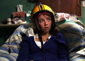
A subject in a God Helmet experiment
It is safe to say that we may never know if our creator resides inside our own minds, in another inaccessible dimension or walks among us. Until then, we can only investigate our own existence as a function of what we can observe or from what we can infer about information we have already obtained. For a more in depth exploration of the creator question, check out Discovery Channel's "Through The Wormole" narrated by Morgan Freeman (just follow the consecutive parts!).
An article about Persinger's God Helmet
The "Through the Wormhole" segment on Persinger's work
Wikipedia articles on the God Helmet and Persinger
Engineering the Blind to See
![]()
Vision is one of the most impressive functions of the human brain. It interprets nothing but electromagnetic waves and paints a glorious picture of our daily existence from the scattered chaotic sea of intertwining light waves that we call home. Many see their vision deteriorate and the world blur as time goes on and these problems can be corrected by optometry, but blindness comes on like a relentless infidel for more than two million people worldwide in the form of retinitis pigmentosa (RP). RP is a heritible genetic disorder that leads to degeneration and loss of function in the retina's photoreceptor cells, and can lead to full blindness in a matter of years. There is no cure or treatment for RP, but new research may change that very soon.
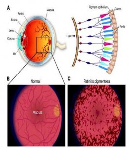
TOP: Macroscopic human eye (left) and rod and cone cell arrangement on the retina (right). BOTTOM: Normal human retina (left) and human retina with retinitis pigmentosa (right)/(Source: Medical-Look.com)
Vision starts in the photoreceptor when light activates rhodopsin, a G-protein coupled receptor pigment consisting of light sensitive opsin and retinal. Light triggers a conformational change in retinal that kick starts a G-coupled visual cascade and the flow of visual information to the brain. In retinitis pigmentosa, rhodopsins in the photoreceptors become insensitive to light starting in the rod cells, and blindness sets in gradually. Rod cells are used in low light and deteriorate first, leading to night blindness, and dysfunction in cone cells used for color vision and acuity sets in until full blindness plagues the individual. Fortunately, a team of French scientists has investigated this degradation and has found a way to combat it by reactivating the photoreceptor cells through genetics. Their study was featured in the July 23rd issue of Science.
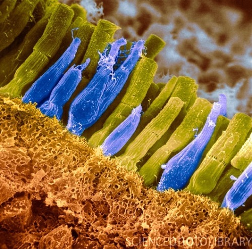
Artificially colored micrograph image of retinal rods (yellow-green) and cones (blue). (Credit: Science Photo Library @ sciencephoto.com)
The scientists isolated an achaebacterial rhodopsin analog called halorhodopsin that functions in the yellow and green wavelength range. They then introduced a halorhodopsin encoding gene into retinitis pigmentosa model mice via a viral vector and also created a control group. In their experiments, it was found that both slow and fast degrading retinal cells in the experimental mice regained their light sensitivity in response to integration of halorhodopsin into their insensitive photoreceptor cells. Electrical responses were recorded from ganglion cells (the third tier cell in the visual cascade) and healthy photoreceptor spikes were observed in response to light stimulation. Most importantly, lateral inhibition (the mechanism by which the brain discriminates edges of objects) was fully preserved, while mono-directional movement was retained. Halorhodopsin mice also performed significantly better than the control RP mice in a battery of visually guided tasks, demonstrating that their photoreceptors had been successfully resensitized by halorhodopsin integration. The scientists also tested the resensitizing ability of halorhodopsin on cultured human retinal cells. They were successful in integrating halorhodopsin into human cells via viral vectors, but could not conduct any clinical trials. However, photoreceptors expressing halorhodopsin demonstrated photocurrents and photovoltages that would be adequate to restore human vision.

Theoretical device that allows a halorhodopsin treated patient to see by projecting patterned light onto the eyes derived from camera input. (Credit: Y. Greenman/Science)
This opens the door to treatment of retinitis pigmentosa in the genes - the same place where it starts. Although halorhodopsin therapy will not fully restore all wavelengths in human vision, it can still serve as a tool to bring restore vision in the blind through optical devices. For example, an RP patient could be treated with halorhodopsin gene thearpy, then outfitted with a device that images the visual field and translates it into halorhodopsin recognizable wavelengths. This light mosaic is then projected onto the patient's eyes and the can "see" what is in front of them. The supplied image of the device is from a perspective article in the beginning of the current issue of Science.
View the full text Science article here (HTML) or here (PDF) and the perspective piece about the article here. Be sure to discuss in the comments!
Sources:
Seeing the Light of Day - Science (Perspective)
Genetic Reactivation of Cone Photoreceptors Restores Responses in Retinitis Pigmentosa - Science (Research Article)
Opening Eyes to Learning Difficulties
![]()
Learning difficulty and disability has long been a problem for many children, parents and school teachers alike. Dysfunctions such as dyslexia and motor disability have hindered the progress of countless adolescents across the country and continue to do so with every passing day. Now, studies have been performed that may centralize learning difficulties to the eye, rather than the brain itself.
Researchers at the Norwegian University of Science and Technology are conducting research that creates a causal link between motor and learning disabilities and dysfunction in visual perception. For example, people who cannot quickly learn a simple motor task such as catching a ball may have difficulty because the cells in their eyes are not perceiving the stimulus properly. The same rings true in individuals with dyslexia - their eyes may not be correctly processing the visual stimuli of words on the page.

Learning disability has long puzzled victims, observers and scientists.
The ocular cells in contest here are deemed "magno cells" and detect rapid movements in our visual field, creating the movie-like perception we experience on a daily basis. Without these, life would look like a disconnected string of frames - much like a comic book. In a test conducted by the researchers, it was found that individuals with difficulty in mathematics also showed difficulty in tracking the randomized movement of a dot on a screen with their eyes, elucidating a link between eye function efficiency, detection of rapid changes in the environment and learning ability.
In a greater context, this finding may have implications in special education and may change the mindset of those working with individuals with additional learning needs. With this new information, learning disability can be combated from the angle of visual field perception. Techniques aiming to strengthen visual perception and eye efficiency (such as eye movement and tracking exercises) could act as a therapy for learning or motor disability previously thought to be localized in the brain itself.
Source: Science Daily via The Norwegian University of Science and Technology
Stressed? It may be in your genes…
![]()
With finals around the corner, the stress factor on campus is bound to rise in the next few weeks. Individual students have their own way of coping with stress, such as TV, video games, music, power naps at Mugar Library (the cubbies are quite comfortable) or a marathon visit to FitRec. Regardless of the method, all aim to reduce the anxiety of coming exams.
Stress also has a neurophysiological effect that decreases rates of neuroplasticity (the ability for the brain to "rewire" itself) in the hippocampus - an area believed to be a center of learning and memory. Additionally, new research suggests that there is a genetic basis for stress effects on the brain connected to a protein called brain derived neurotrophic factor (BDNF). In a joint study conducted by Rockefeller University and Weill Cornell Medical College researchers, it was found that mice with inadequate BDNF expression had brains that looked similar to those of mice exposed to chronic stress over a long period of time. These mice were not exposed to chronic stress, yet they still showed decreased rates of neuroplasticity in the hippocampus - a characteristic of stressed brains.
It seems that stress is a product of our environment as well as our genes. At this point, research on BDNF is just beginning. However, this research might open doors to the treatment of chronic stress disorders on the genetic and physiological level without the use of controversial psychotropic drugs.
Read the full article over at Science Daily for the whole scoop.
Gene That Changes the Brain’s Response to Stress Identified
via Science Daily
