Transcranial retinal imaging
Widefield human retinal imaging is typically performed in a reflection geometry, where light is delivered through the pupil and images are formed from the light reflected back from the retina. In this configuration, artifacts caused by retinal surface back-reflections are often encountered, which complicate quantitative interpretation of the reflection images.
We have developed an alternative widefield retinal imaging technique based on near-infrared light delivered transcranially through the subject’s temple. The light diffuses through the bone and illuminates the retina not from the front, as in standard techniques, but rather mostly from the back. As such, images are formed from light transmitted through the retina rather than reflected from the retina. This unique transmission geometry simplifies absorption pathlength considerations and enables flash-free, non-mydriatic imaging as deep as the choroid. Multiple wavelengths may be used enabling measurement of blood oxygen saturation in retinal vessels.
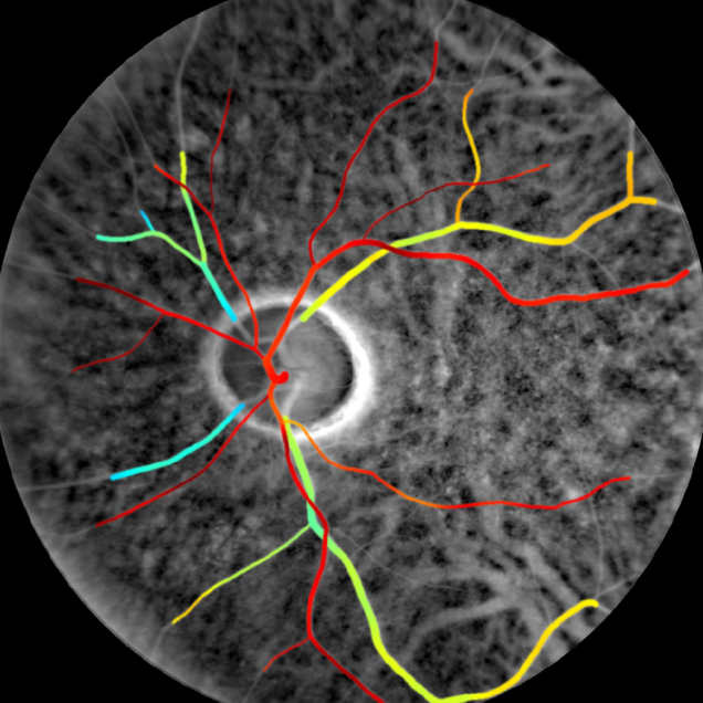
- T. D. Weber and J. Mertz, “Non-mydriatic chorioretinal imaging in a transmission geometry and application to retinal oximetry”, Biomed. Opt. Express 9, 3867-3882 (2018). link