Oblique back-illumination microscopy
Phase contrast microscopy provides exquisite high-resolution images of sample morphology, without the use of sample labeling. However standard phase contrast techniques, like differential interference contrast (DIC), only work in the transmission direction and thus cannot be used when imaging thick samples. For this reason, standard phase contrast techniques have not made a great impact in in-vivo biomedical or clinical imaging applications.
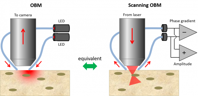
We have developed a new technique, called Oblique Back-illumination Microscopy (OBM) that provides microscopic resolution DIC-like images of sub-surface sample morphology even in very thick tissue. In addition, OBM provides simultaneous amplitude imaging by a method of shadowcasting. Because OBM works in a reflected light geometry, it is amenable to endoscopy applications.
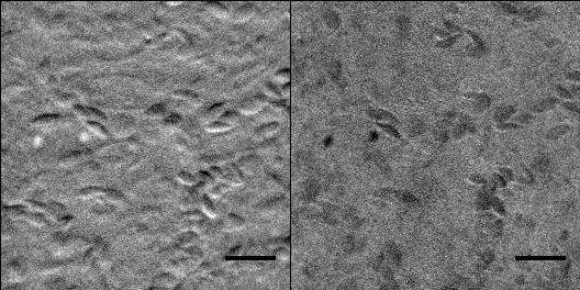
Two configurations of OBM are equivalent by reciprocity. The first is a camera-based configuration that uses standard widefield detection optics, but offset illumiantion optics. In the case of an endoscope, some extra relay optics is introduced in the detection path, such as an imaging fiber bundle. The second configuration (sOBM) is a laser-scanning-based configuration equipped with offset detection. This second configuration has the advantage that it can be combined with standard laser-scanning microscopes such as two-photon or coherent R15. Single-shot OBM video of chick embryo vasculature/OBM produces phase-gradient (left) and amplitude (right) contrast simultaneously. Single-shot operation is achieved by using two different illumination colors. Acquisition is video rate.aman microscopes, yield multi-contrast images that are automatically co-registered.
- S. Xiao, S. Zheng, J. Mertz, “High-speed multifocus phase imaging in thick tissue”, Biomed. Opt. Exp. 12, 5782-5792 (2021). link
- C. Ba, J.-M. Tsang, J. Mertz, “Fast hyperspectral phase and amplitude imaging in scattering tissue”, Opt. Lett. 43, 2058-2061 (2018). link
- C. Ba, M. Palmiere, J. Ritt, J. Mertz, “Dual-modality endomicroscopy with co-registered fluorescence and phase contrast”, Biomed. Opt. Express 7, 3403-3411 (2016). link
- J. Li, T. G. Bifano, J. Mertz, “Widefield fluorescence microscopy with sensor-based conjugate adaptive optics using oblique back illumination”, J. Biomed. Opt. 21,121504 (2016). link
- J. Mertz, A. Gasecka, A. Daradich, I. Davison, D. Coté, “Phase gradient contrast in thick tissue with a scanning microscope”, Biomed. Opt. Express 5, 407-416 (2014). link
- J.-D. Giese, T. N. Ford, J. Mertz, “Fast volumetric phase-gradient imaging in thick samples”, Opt. Express 22, 1152-62 (2014). link
- T. N. Ford, J. Mertz, “Video-rate imaging of microcirculation with single-exposure oblique back-illumination microscopy”, J. Biomed Opt. 18, 066007 (2013). link
- T. N. Ford, K. K. Chu, J. Mertz, “Phase-gradient microscopy in thick tissue with oblique back-illumination”, Nat. Meth. 9, 1195-1197 (2012). link