Research
Studies Related to Spectroscopy for Detecting Head & Neck Cancer
The ability to detect cancer cells in head and neck tissue is critical for diagnosis and treatment of head and neck cancer (HNC). Although tissue biopsy and histological analysis is the gold standard for detecting HNC cells, it is invasive, time-consuming, and expensive, requiring the work of several medical specialists.
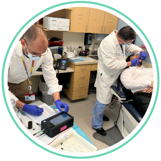
Spectroscopy – the study of how light interacts with tissues – offers a relatively inexpensive, non-invasive alternative to cancer cell detection. The premise of this approach is that light exhibits different refraction patterns when it interacts with cancer cells instead of healthy cells due to differences in cellular micro-architecture, or in other words: light that has bounced off of cancer cells looks different from light that has bounced off of healthy cells.
Dr. Irving Bigio and the Biomedical Optics Lab at Boston University have pioneered the application of elastic scattering spectroscopy (ESS) for cancer cell detection using devices made up of a lamp, to generate light; a probe, to direct light onto and collect light from tissue; a spectrometer, to generate light spectral data; and a computer, to operate the device and record light spectra. ESS algorithms developed using artificial intelligence and machine learning techniques can distinguish light that has interacted with cancer cells from light that has interacted with healthy cells. Each ESS measurement takes less than a second to capture, record and analyze, requiring no special dyes, lighting, pathology expertise or highly technical equipment. The Biomedical Optics Lab and the OTO-COATI Lab have teamed up to develop ESS algorithms that could improve the diagnosis and treatment of head & neck cancer.
Validation of Elastic Scattering Spectroscopy for Intra-operative Margin Guidance during Oral Cancer Resection
Contributing Lab Faculty: Gregory Grillone, MD (PI) & Gintas Krisciunas, PhD, MPH, MA
Contributing Lab Staff & Students: Lucas Berry, Devin Lucas, Nikhita Santebennur & Gabriele Spokas
Collaborating Investigators: Irving Bigio, PhD – Boston University; John Gooey, MBBS – VA Boston Healthcare System; Miriam O’Leary, MD – Tufts Medical Center
Funding Source: NIDCR/NIH (1R01DE029862)
Detecting Laryngeal Cancer by Applying Light Scattering Spectroscopy to the Oral Cavity – A Pilot Study
Contributing Lab Faculty: Gregory Grillone, MD (PI), Gintas Krisciunas PhD, MPH, MA, & Lauren Tracy, MD
Contributing Lab Staff & Students: Lucas Berry, Gabriele Spokas, & Mitali Sakharkar
Collaborating Investigators: Irving Bigio, PhD – Boston University
Leukoplakia (abnormal white spots) of the larynx (voice box) is a serious finding given that it can transform into laryngeal cancer. ENT specialists monitor patients with laryngeal leukoplakia by examining them with a laryngoscope (lighted tube), and taking samples of the leukoplakic tissue to send to pathologists, who test it for cancer. This monitoring process is both costly and burdensome for the patient, requiring frequent visits to the ENT over several years. ESS has the potential to lessen the burden of this process by providing a faster, less invasive method of assessing laryngeal cancer risk that could be performed by non-specialist providers. Based on the concept of field cancerization, which suggests that oral cavity (mouth) tissue of patients with benign laryngeal leukoplakia is substantially different from oral cavity tissue of patients with malignant laryngeal cancer, the present pilot study aims to assess the diagnostic potential of ESS for laryngeal leukoplakia by collecting ESS measures from patients with benign laryngeal leukoplakia and patients with malignant laryngeal cancer.
Characterization of Oral Cavity Sub-sites via Elastic Scattering Spectroscopy
Contributing Lab Faculty: Gregory Grillone, MD & Gintas Krisciunas PhD, MPH, MA
Contributing Lab Staff & Students: Gabriele Spokas, & Mitali Sakharkar (PI)
Collaborating Investigators: Irving Bigio, PhD – Boston University
Although ESS has the potential for several diagnostic applications based on measures of the oral cavity, its unknown whether this potential could vary by anatomic site. For example, an ESS measure of the tongue may be different from an ESS measure of the gum, and that difference might affect the accuracy of ESS algorithms. The present study aims to investigate this possibility by collecting ESS measures from several different anatomic sites within the oral cavities of healthy individuals. The ESS data generated from this study could be used to refine ESS diagnostic algorithms.
Studies Related to Acute Respiratory Failure
Acute respiratory failure (ARF) occurs when a patient’s medical condition leaves them unable to breathe or protect their airway on their own, requiring a mechanical ventilator to breathe for them via an endotracheal tube (ETT) or tracheostomy (port in the neck/throat). Intubation – the process of inserting an ETT into a patient’s airway – can cause damage to the airway and adjacent anatomical structures. Because of this, ARF survivors are susceptible to severe and potentially life-threatening complications after the tube is removed (post-extubation) like stridor, laryngeal edema, dysphagia, and aspiration, which can lead to adverse outcomes including pneumonia, re-intubation, prolonged hospital stay, and/or death.
The OTO-COATI lab is a part of a nation-wide consortium, led by Dr. Marc Moss at the University of Colorado, which studies complications related to ARF. This consortium elucidates pertinent findings by collecting data from ARF patients’ post-extubation swallow screen results, results of fiberoptic endoscopic examinations of swallow (FEES), and medical records generated during stays in intensive care units (ICUs). The goal driving the consortium’s past and future findings is to develop robust assessments and interventions that will lessen the frequency and burden of complications associated with ARF survivorship.
Aspiration in Acute Respiratory Failure Survivors
Contributing Lab Faculty: Gintas Krisciunas, PhD, MPH, MA (PI)
Contributing Lab Staff & Students: Devin Lucas
Collaborating Investigators: Nicholas Hill, MD – Tufts University; Joseph Levitt, MD MS – Stanford University; Marc Moss, MD – University of Colorado Denver; Jonathan Siner, MD – Yale University
Funding Source: NINR/NIH (1R01NR019989)
Characterizing Post-extubation Laryngeal Edema in the Presence of Aspiration
Contributing Lab Faculty: Gintas Krisciunas, PhD, MPH, MA (PI)
Contributing Lab Staff & Students: Devin Lucas & Mitali Sakharkar
Collaborating Investigators: Marc Moss, MD – University of Colorado Denver
While both laryngeal edema and aspiration commonly occur in ARF survivors post-extubation, the correlation between the two events is unknown. The present study aims to investigate the correlation between the two events using data collected in a previous study.
Studies Related to Dysphagia Outcomes & Assessment
Dysphagia, or impairment of swallowing, can arise as many different presentations and severities, ranging from inconvenient to life-threatening. Dysphagia is primarily managed by speech language pathologists (SLPs). Instrumental exams, including the fiberoptic endoscopic exam of swallow (FEES) and video fluoroscopic swallow study (VFSS), are considered the gold standard diagnostic assessments of dysphagia.
There are few standardized guidelines for evaluating and treating dysphagia, making research on pertinent clinical outcomes and development of novel assessment tools necessary. The OTO-COATI Lab works with expert SLPs and dysphagia researchers from around the world to enhance and elucidate relevant diagnostic capabilities and clinical outcomes.
Validation of the Boston Residue And Clearance Scale (BRACS)
Contributing Lab Faculty: Gintas Krisciunas, PhD, MPH, MA (PI)
Contributing Lab Staff & Students: Lucas Berry, Arpita Edke, Winston Liu, & Mitali Sakharkar
Funding Source: BUMC CTSI
Reliability and Validity of Existing Scales to Measure the Residue Problem
Contributing Lab Faculty: Gintas Krisciunas, PhD, MPH, MA (PI)
Contributing Lab Staff & Students: Lucas Berry
Funding Source: BUMC CTSI
Addressing the same problem as the BRACS study (above), the present study aims to evaluate the reliability and validity of generic scales commonly used to assess the residue problem during FEES exams. This aim will be achieved using data collected from SLPs around the world as they assessed clips of FEES exams representing the broad spectrum of residue problem presentations.
Variation in Dysphagia Evaluation Practices
Contributing Lab Faculty: Gintas Krisciunas, PhD, MPH, MA (PI)
Contributing Lab Staff & Students: Lucas Berry, Arpita Edke, Veronica Han, & Gabriele Spokas
Funding Source: BUMC CTSI
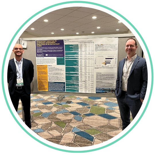
Variation in care can occur in the absence of clear, empirically supported clinical guidelines that are known to promote optimal patient outcomes and appropriate use of health care resources. Given a lack of evidence-based algorithms for choosing the type or number of dysphagia associated assessments, the present study seeks to use retrospective medical record data to determine whether there was variation in administration of instrumental dysphagia assessments (e.g. FEES, VFSS) or patient reported outcome measures (PROMs) related to dysphagia in an outpatient clinic.
Studies Related to Biofeedback for Voice Disorders
When a person’s perilaryngeal muscles (muscles connected to the voice box) are over-active, they have vocal hyperfunction (VH). VH is associated with the most common types of voice disorders, including those that happen after repeated trauma to the vocal cord tissue (e.g. nodules and polyps) and vocal deterioration that occurs without a clear medical cause (muscle tension dysphonia).
The OTO-COATI lab studies VH in collaboration with the Vocal Hyperfunction Clinical Research Center (VHCRC) at Massachusetts General Hospital, which is led by Robert Hillman, PhD, CCC-SLP, research director and co-director of the Mass General Voice Center. The VHCRC is is an interdisciplinary, multi-institutional research program which integrates many types of data to attain a better understanding of the multiple causative factors and disordered physiological processes associated with VH in order to develop new, more effective methods for prevention, diagnosis and treatment.

