Heaphy Lab
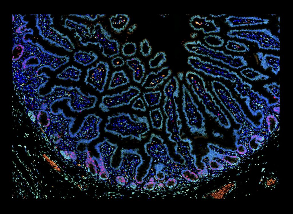
Ki67 in small intestine
Ki67 (red) and lamin A/C (green) IF
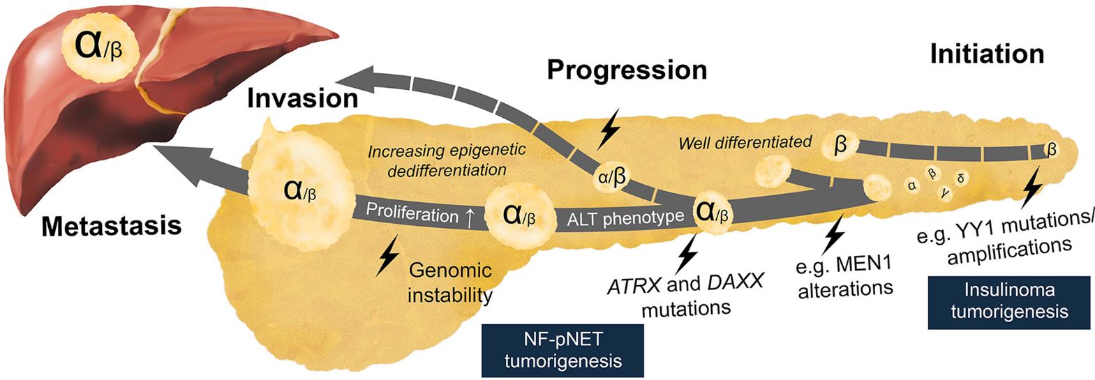
Proposed PanNET model
Multistep tumorigenesis of sporadic non-functional PanNETs
Telo-CISH
Telo-CISH multiplex with basal-specific cytokeratin facilitates easy identification of short telomeres
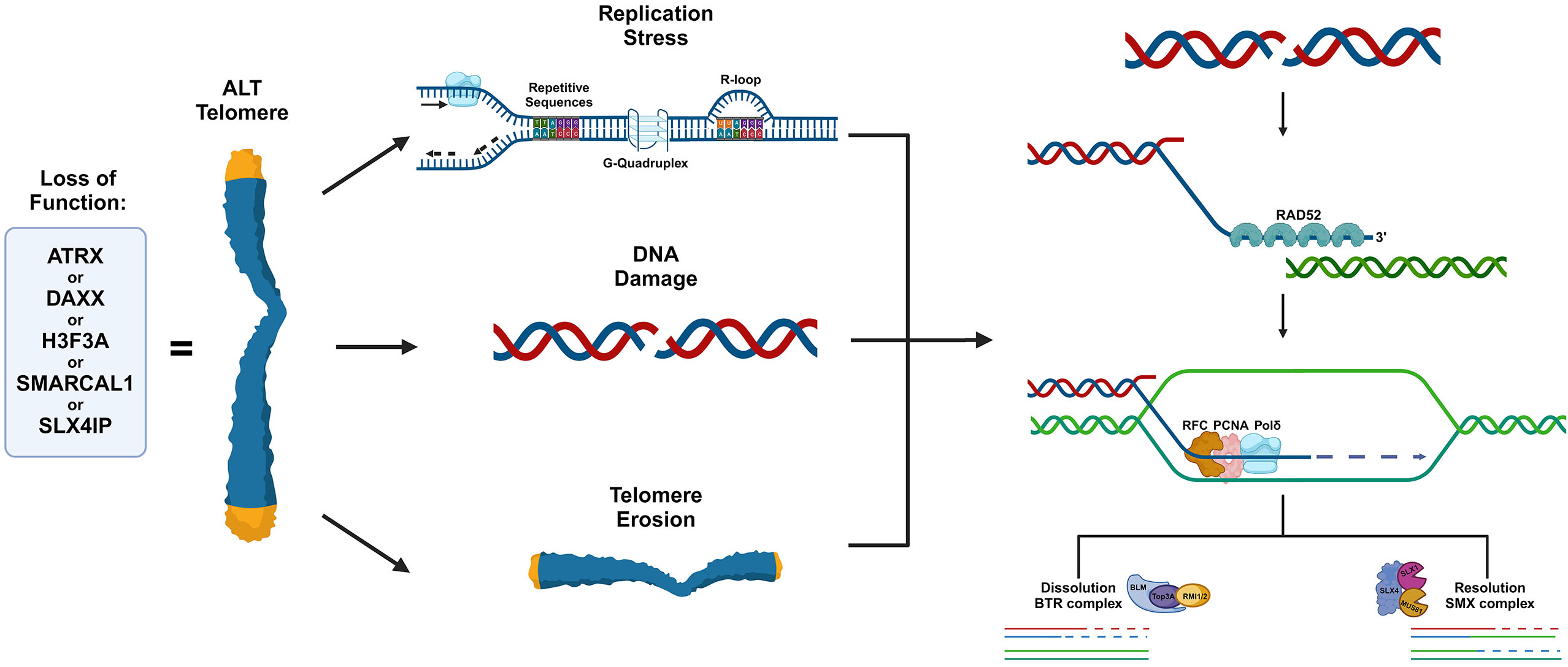
ALT mechanism
Break-induced replication mediates the extension of ALT telomeres

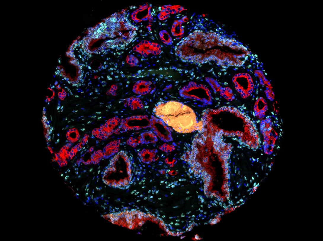
Lunaphore COMET
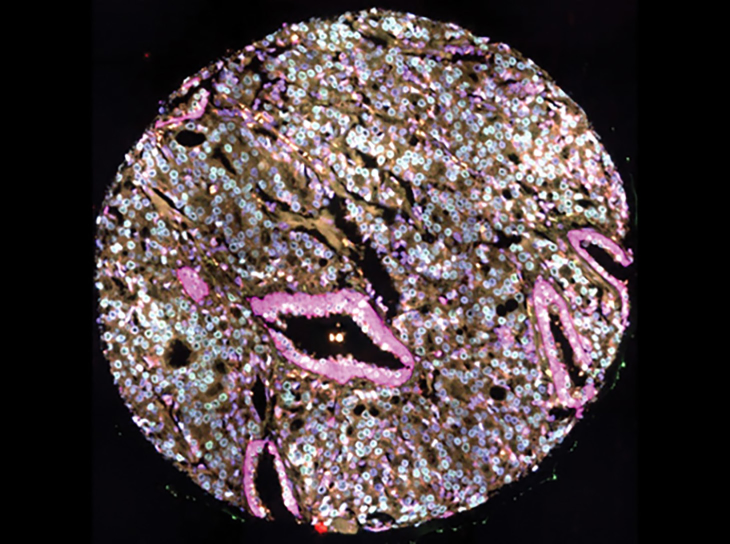
mIF (pan-CK & CD56) in PanNETs
Breast TDLU
Telomere and centromere-specific FISH with costaining for smooth muscle actin (magenta) and Ki67 (turquoise)
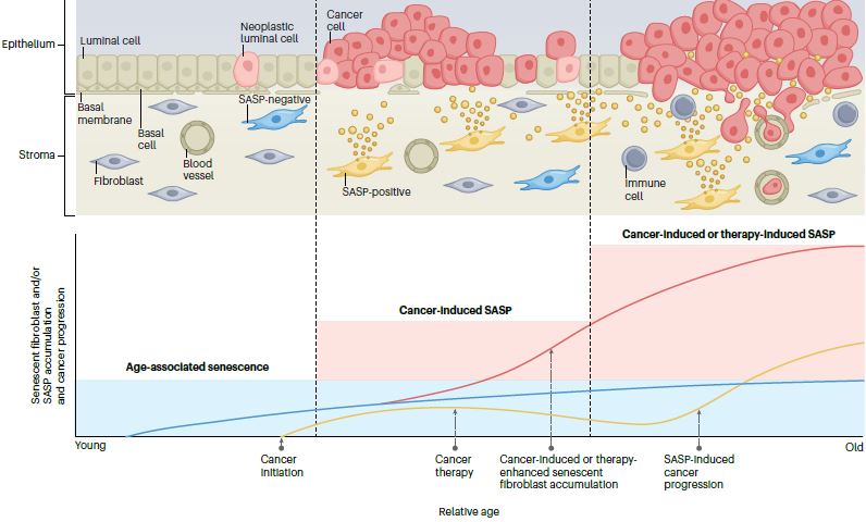
Senescent fibroblasts
Senescent fibroblasts can accumulate and produce senescence-associated secretory phenotype (SASP).
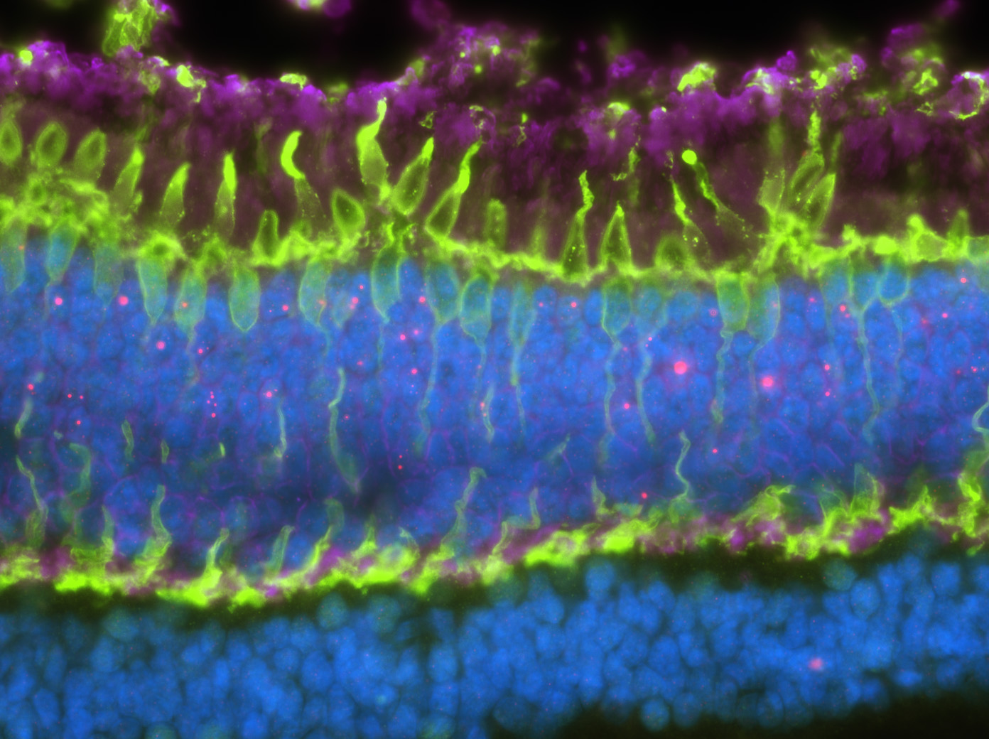
Photoreceptors
Telomere-specific FISH with colabeling for cone nuclei and processes (green) and rod nuclei (magenta)
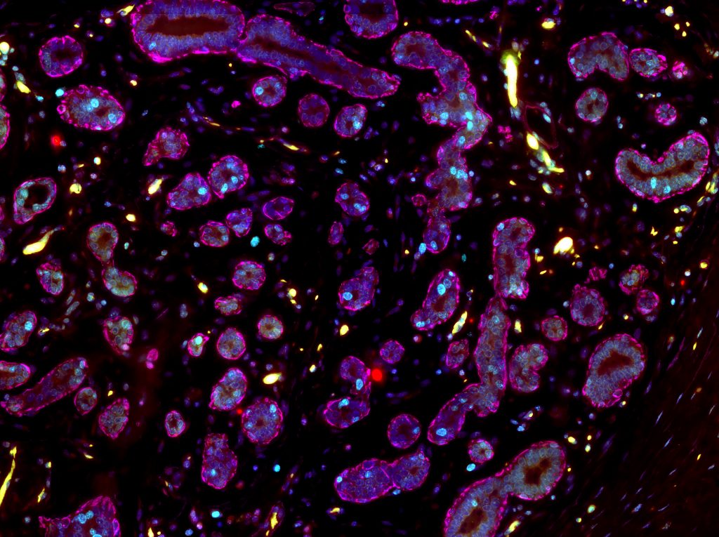
Prostate Cancer TMA
Telomere-specific FISH with costaining for a basal-specific cytokeratin (magenta) as well as NKX3.1 and FOXA1 (green)
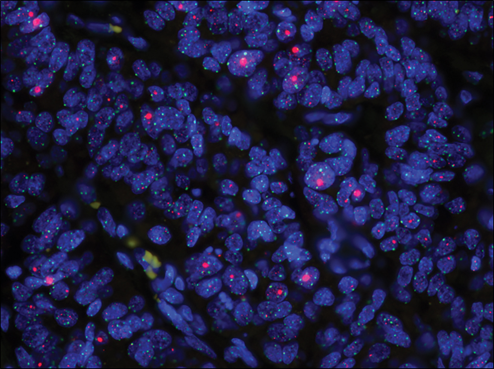
ALT+ PanNET
Telomere-specific (red) and centromere-specific (green) FISH denotes the presence of ALT
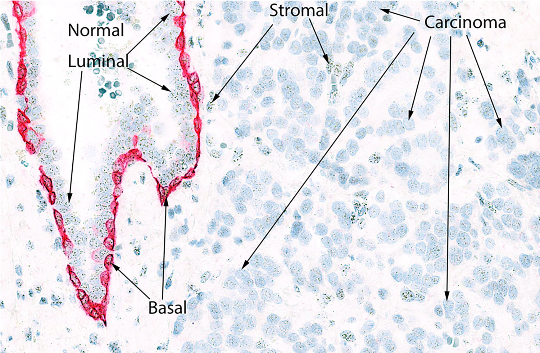
Lamin A/C in PrCa
Costaining for a lamin A/C (green) and cytokeratin 8 in a prostate cancer
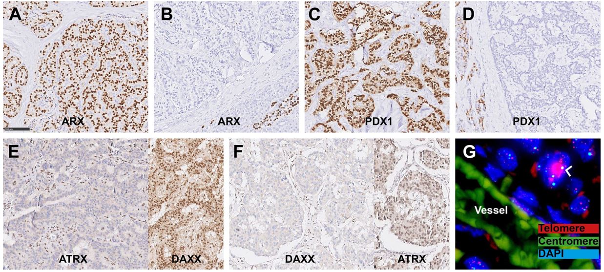
PanNET biomarkers
Established tissue-based prognostic biomarkers for non-functional pancreatic neuroendocrine tumors.
HEAPHY LAB MISSION
The Heaphy Lab applies an integrated, multidisciplinary approach, which combines tissue-level analysis, cell-based modeling, and molecular and spatial profiling technologies, to define how telomere dysfunction drives the initiation and progression of cancer. Our work spans pancreatic neuroendocrine tumors, prostate cancer, breast cancer, sarcomas, and gliomas.
We aim to translate fundamental discoveries in tumor and tumor microenvironment (TME) biology into clinically actionable biomarkers and strategies that improve cancer risk stratification, prognostication, and therapeutic decision-making.
Equally central to our mission is fostering a rigorous, collaborative, and inclusive laboratory culture. We are deeply committed to mentoring and supporting the professional and personal development of every lab member, empowering the pursuit of impactful, independent, and team-driven research.