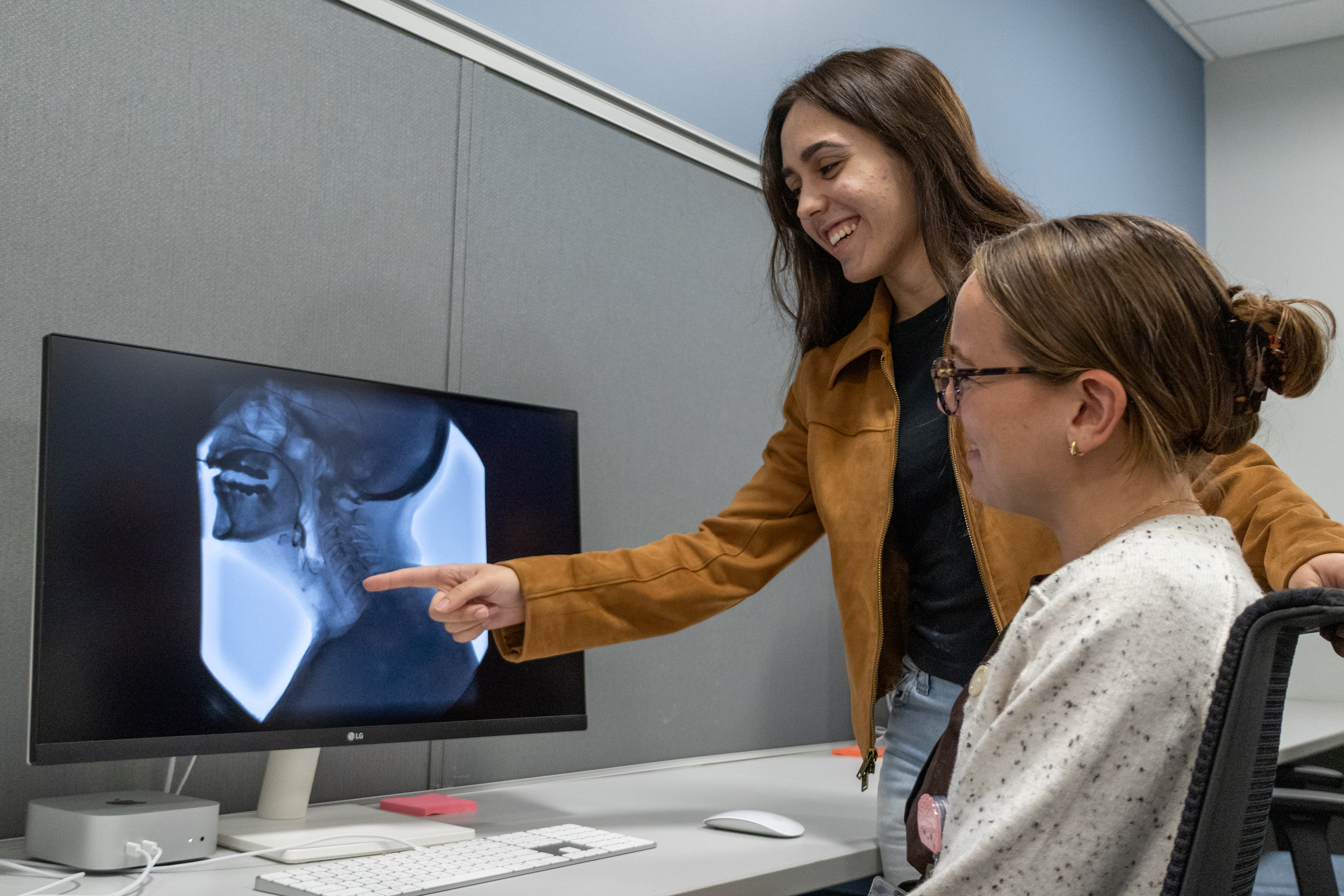
The Airway Protection Research Lab is dedicated to advancing our understanding of cough and swallowing disorders so that we can translate these findings into novel, evidence-based treatments. Our overarching goal is to improve health outcomes and quality of life for patients and care partners. To accomplish these goals, we use a range of clinical and research tools, including:

Videofluoroscopic Swallowing Imaging

Still image from x-ray video of swallowingVideofluoroscopy is one of the most common tools used to evaluate swallowing. During this test, a person eats and drinks small amounts of food and liquid mixed with a contrast material. An x-ray video is taken while the person swallows, allowing us to watch how food and liquid move through the throat in real time. This test allows us to visualize anatomy, physiology, and bolus movement to evaluate swallowing function and determine the effectiveness of strategies and interventions that improve safety and efficiency. Although videofluoroscopy involves a small amount of radiation, the risk is very low. The radiation exposure from this test is much lower than other types of imaging exams. In fact, it would take more than 40 videofluoroscopy studies to exceed the radiation exposure from a routine chest CT scan.


Cough and Respiratory Measures

Chart showing coughing and breathing measurements collected using spirometry, respiratory plethysmography, and handheld peak flow meters

We measure coughing and breathing using gold-standard tools such as spirometry, respiratory plethysmography, and handheld peak flow meters. These methods allow us to measure different aspects of coughing and breathing function, such as lung volume or the strength and timing of a cough. We also assess cough sensitivity using small amounts of safe sensory stimuli to understand how the body responds to airway stimulation.

Brain Imaging

Brain imaging using infrared spectroscopy (fNIRS)Functional near-infrared spectroscopy (fNIRS) is a safe and non-invasive way to measure brain activity. It uses a soft cap worn on the head and works well during tasks that involve some degree of movement, such as swallowing or coughing. We use fNIRS to study how the brain controls airway protection and how treatment improves brain function. We work closely with collaborators at the BU Neurophotonics Center to use state-of-the-art brain imaging tools to answer these and other research questions.


Meta-Science and Causal Modeling

Answering complex questions about airway protection requires science that is rigorous, transparent, and reproducible. To meet these standards, our lab emphasizes areas like causal inference, statistical modeling, and clinical trial design. We collaborate closely with experts across these disciplines to rigorously address questions such as how swallowing and cough function change over time and which physiologic, cognitive, or treatment-related factors drive improvement.

These analytic approaches allow us to move beyond simple correlations and instead interrogate casual mechanisms that meaningfully inform clinical practice. Our lab is equally committed to meta-science, with the goal of improving how our research is designed, conducted, analyzed, and reported. This includes strengthening the transparency of our work through open science practices, reducing sources of bias, enhancing analytic rigor, and promoting computational reproducibility.

Through these efforts, we aim to foster a scientific environment that is open, diverse, and reproducible, ultimately maximizing the impact of our research for patients, clinicians, and care partners.


Want more information? 
Email us at aprlab@bu.edu

Borders Airway Lab Logo