Brain-synching: What Happens When You Converse with Other People
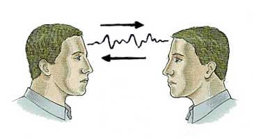
Over-used expressions like “on the same page” or “same wavelength” may actually have some physiological truth behind them. When two people are having a conversation or listening to the same story, it makes sense that they’d be using similar parts of their brain, but the question is just how similar this activation is. Drexel and Princeton Universities teamed up recently to further explore this issue using a newer technology, hoping to prove its efficacy.
Using functional near-infrared spectroscopy (fNIRS), a functional brain imaging technique, researchers sought to find out what happens when two people communicate and how to possibly improve face to face communication. In this study, experimental subjects wore an fNIRS headband which measured their neural activity while they engaged in conversation with one another. This in itself is pretty great as other imaging techniques like fMRI that measure blood flow to brain regions require people to lie down in a noisy machine, which is not at all conducive to personal conversation.
During the experiment, subjects listened to a story in their native language while their futuristic headbands measured activity in prefrontal and parietal areas. These regions were targeted because they’re largely responsible for higher order processing involved with relating to others, an important piece of any communicative effort. When they examined the recordings, the researchers saw that brain activity of the listener heavily resembled that of the speaker after a delay. This copy-cat effect, however, was not observed when subjects didn’t understand the communicator, for example when the speaker only communicated in Turkish but the listener was only fluent in English.
With the results from fNIRS, the experimenters found that the fNIRS recordings correlated quite closely with fMRI results of a similar experiment. This is a pretty big deal since it confirms that fNIRS is a legitimate functional imaging technique that could open the door to a brand new wave of experiments involving communication. fNIRS will prove a useful tool in the future, especially to decode the issue of “brain-synching” during conversation.
~ Jackie Rocheleau
Sources:
Measuring speaker-listener neural coupling with functioning near infrared spectroscopy
Brain imaging headband measures how our minds align when we communicate
Image Source:
https://cdn-images-1.medium.com/max/800/0*BZrVsheqbGZyc954.jpeg
Science of Happiness: What Makes Us Truly Happy?

Happiness, by definition, is often the feeling of contentment or pleasure in doing something you like. However, the formal definition of happiness and what it truly is may differ. In one study, the Harvard Department of Psychology tried to determine the role of morality in happiness. The participants were given an example of a hypothetical person named “Tom", who rarely felt sad or lonely. This is because he felt satisfied by stealing from students and reselling the items he stole to buy alcohol. Most of the subjects agreed that Tom is satisfied, but not exactly happy. The way they attributed this was that one has to be good to be happy, and therefore morality plays a role in happiness. This was surprising because Tom was clearly happy with his decisions but was deemed unhappy by people who have never even known this hypothetical character. So what exactly makes us happy?
In another study conducted by Dr. Kringelbach and Dr. Berridge, they researched and found the exact neuroscience behind happiness and pleasure. They started by comparing the correlation between happiness and hedonia (pleasure) and the correlation between happiness and eudaimonia (a life well lived). Through this, they found that most people associated happiness more strongly with hedonia, which explains why Tom might feel happy about his actions. They then identified the hedonic hotspots in the brain, which include centers that produce “neurochemical modulators” and enhance a liking reaction. This pleasure that is experienced would then be translated into a motivational process that would lead to wanting, as it would increase dopamine (the neurochemical for pleasure) in the brain. By enhancing this connection between what makes you happy and the chemicals in your brain, you involuntarily strengthen actions that create pleasure through constant usage. An important note from this study is that “all pleasures seem to involve the same hedonic brain systems, even when linked to anticipation and memory.” This would lead individuals without certain “higher-order pleasures (such as monetary, artistic, musical, altruistic and transcendent pleasures)” to search for other ways to satisfy that biological need.
So how can we search for or create happiness? Dan Gilbert explains in a TED talk that happiness can be synthesized. He brings up the concept of a psychological immune system- a way for you to feel better about the world you live in. This may come in the form of morality, such as when the students from the initial study thought that the hypothetical person, Tom, could only be happy by being moral. It could also be the way Tom finds happiness through immoral actions. Gilbert explains that “the freedom of choice is an enemy to synthetic happiness.” Having a choice in what makes you happy creates a negative effect while being stuck with a choice makes you grow to like what you choose. Once the decision becomes a choice, the indecisiveness may become detrimental to your happiness. In order to be truly happy, you must simply stick to a choice and be happy about it.
If you have time, check out the TED talk below, as it is truly a great watch.
~Albert Wang
Sources:
TED talk: What makes you happy?
What Does It Take to Be Truly Happy
The Neuroscience of Happiness and Pleasure
Image Source:
http://www.marcandangel.com/images/9-not-need-happy.jpg
Sleep & Memory Consolidation

As important as it is to be productive and live a balanced life, it seems like for a great deal of people, students in particular, health comes second to term papers. I suppose it’s an occupational hazard, but it’s pretty interesting to think that despite the incredibly adverse affects on our intellect, sleep is the first healthy habit to go.
Most people have figured out on their own that we need sleep (or caffeine) to recharge and start the next day energized, but they seem to ignore the other equally important benefits. The second most known benefit of sleep is likely that it helps our immune systems function, which, while obviously important, can also be supplemented to an extent with vitamins and healthy eating. However, the one function of sleep that we absolutely cannot replace is memory consolidation.
Basically, when you encounter or experience something important, an area of the brain called the hippocampus becomes active and works to hold that event in your short term memory. In order for these short term memories to become long term, and thus less easily forgotten, the brain needs to consolidate the information and store it outside of the hippocampus. This process involves the formation, breakdown, and reformation of synapses (connections between neurons) throughout the cortex. It turns out that one of the best things you can do to ensure that this process goes smoothly is sleep.
When you sleep, your brain “downscales” the unimportant activity of irrelevant synapses and “upscales,” or increases activity of, important synapses. A four-year-long study recently published physical proof of this phenomenon in the form of actual pictures of synapse activity in mice: “they found that a few hours of sleep led on average to an 18 percent decrease in the size of the synapses.” (Neuroscience News). Similar findings have been found in people too.
Ironically, students give up a lot of sleep in the name of studying information they’ll never fully retain because of that lack of sleep.
~ Jackie Rocheleau
Sources:
How the Brain Resets During Sleep
Image Source:
https://www.michigandaily.com/sites/default/files/leg/imagecache/fullnode/zzzz.jpg
Why Multitasking Isn’t a Good Idea

In theory, multitasking sounds efficient. Why perform different tasks separately when you can perform them simultaneously and save time, right? In practice, however, multitasking is not as efficient as it may seem. In fact, people are terrible multitaskers; most attempts at multitasking usually only result in one’s attention switching back and forth between tasks, which exhausts the brain of oxygenated glucose. Because of how tiring this switching is, it becomes harder to successfully focus on a single task.
Multitasking is more than just exhausting, though. In a study performed at Institut National de la Santé et de la Recherche Médicale, scientists found that the brain’s prefrontal cortex, involved with attention, has a left and right side. These sides cooperate when people focus on a single task and work independently when they perform two tasks at once. Participants were asked to perform two tasks simultaneously – when scientists told the participants that they would receive a larger reward for accurately completing one of the tasks, fMRI showed increased neural activity in only one side of the prefrontal cortex. When the larger reward became associated instead with the other task, the other side of the prefrontal cortex showed more neural activity, suggesting that the brain splits in half when there are two simultaneous goals. Additionally, when asked to perform a third task, participants repeatedly forgot one of the three tasks they were asked to perform and were three times as likely to make errors as they had when trying to perform two tasks. The study shows that trying to perform more than two tasks at the same time is more challenging because the brain only has two frontal lobes that can attend to the tasks. While some tasks may be difficult to perform simultaneously, others seem much easier. For instance, reading and eating at the same time is easier than reading and driving because eating demands less engagement from the prefrontal cortex than driving.
Since multitasking generally increases the likelihood of error and is mentally exhausting, how else can a person cope with a busy schedule? Instead of multitasking, scientists recommend taking breaks every two hours, and devoting different activities to specific timeslots, such as only using social media in the morning and midday. As a result of less distraction, productivity will increase and stress will decrease.
~ Nathaniel Meshberg
Sources:
https://qz.com/722661/neuroscientists-say-multitasking-literally-drains-the-energy-reserves-of-your-brain/
http://www.brainfacts.org/sensing-thinking-behaving/awareness-and-attention/articles/2013/the-multitasking-mind/
Image Source:
https://timemanagementninja.com/wp-content/uploads/2013/09/Triple-Tasking.jpg
Study, Sleep, Repeat

This timeline will be familiar to those of you who have experienced an all-nighter. During the first 16 hours of day 1, you feel normal. Your attention span and working memory have not yet been affected. Then, around hour 17, you enter your “biological night time.” The hormone melatonin, which circulates from your brain to your body, reaches a peak level that signals to your body that it is night-time. This is when your performance rapidly deteriorates and reaches a minimum around 6 to 8am the next morning. While your performance may improve throughout the following day, it will remain below that of day one until you get a decent amount of sleep. This timeline of your performance is regulated by your internal biological time of day; it is not a linear deterioration based on the number of hours you have been awake.
A team of researchers used functional magnetic resonance imaging (fMRI), a noninvasive technique used to measure and map brain activity by detecting changes in blood oxygenation and flow, to scan the brains of 33 people who were sleep deprived over two days and following a period of recovery sleep. The participants’ levels of melatonin were also measured to determine each person’s internal biological time. Brain images were taken during a reaction time task, sleep deprivation in the evening and morning when performance undergoes rapid changes, and after recovery sleep. The results showed some variation in the timing of the 24-hour circadian rhythm that is followed by some brain regions, including subcortical areas such as the thalamus, which is responsible for relaying sensory information from receptors in the body to the cerebral cortex. The frontal brain regions showed a decrease in activity during sleep deprivation and a return to regular levels of activity after recovery sleep. The effects of sleep deprivation were also evident in participants’ performances in simple reaction time tasks.
While sleep deprivation affects various brain regions differently, its effects are pervasive. Hence, you should try to sleep between study sessions so your brain has a chance to consolidate the information you studied, and you can be at your top performance level to continue studying or to take your final the next day.
~ Sophia Hon
Sources:
http://science.sciencemag.org/content/353/6300/687.full
http://www.yourhormones.info/hormones/melatonin.aspx
Image Source:
https://metrouk2.files.wordpress.com/2015/01/sleepygif.gif?w=620&h=265&crop=1
How Artificial Intelligence is Beginning to Sense and Perceive
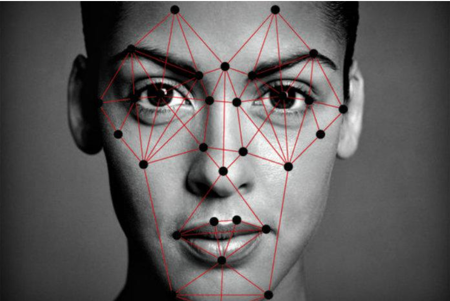
Visual and auditory neuroscience has been used more frequently in the new age of technology. We can see it being used when Facebook automatically identifies our faces in tagged photos or when Siri finally figures out that we want the weather for today and not asking to call your mom. Artificial intelligence is now crossing over onto various different biological sciences, but it’s the scientists and engineers that need to figure out how to transfer all of the information on how natural sounds and visuals get transferred to a computer and how to adapt from that information.
Visual neuroscience’s main focus nowadays is on face processing, or face recognition. The part of the brain responsible for facial recognition is the occipito-temporal region, the right middle fusiform gyrus specifically. Facial recognition is said to involve several stages that range from perceiving basic stimuli to deriving details from said basic stimuli. New research has shown that facial recognition is a very connected brain mechanism that can be taught to those who have experienced a brain injury.
By applying this basic biological science to artificial intelligence, many researchers have been able to further progress the technology surrounding perception – like audial and visual. MIT has recently created a “machine-learning system [that] spontaneously reproduces aspect of human neurology,” meaning that this computational model they have created learns in the same way the brain learns. Apparently this detail wasn’t knowingly built into the system, as it suddenly showed up during the training process. MIT’s Tomaso Poggio developed the system to train itself to recognize certain faces in certain directions. The way they figured out that the system induced another processing step was when a certain face was rotated a certain degree regardless of the direction of the face. Another MIT lab, Computer Science and Artificial Intelligence Laboratory (CSAIL), is attempting to create another machine-learning system that can identify and learn from said identification natural sounds or background noise like crowds cheering or waves crashing. What makes this machine-learning system different from its predecessors is the fact that it does not require hand-annotated data when training, and instead researchers use videos to find the correlations between visuals and sounds. Carl Vondrick, graduate student of the MIT lab, describes it as “the natural synchronization between vision and sound.”
The sciences between artificial intelligence and biology are growing closer and closer together as this era of technology progresses furthermore. It was only a few years ago that facial recognition software became wholly available to the public, and so with further advancement in technology, who knows what one might expect with artificial intelligence.
~Cindy Wu
Sources:
http://neurosciencenews.com/machine-learning-facial-recognition-5654/
http://neurosciencenews.com/video-sound-machine-learning-5665/
https://www.sciencedaily.com/releases/1999/06/990624080203.htm
Why Are You Copying Me?

Ever notice someone, friend or stranger, subconsciously mimic your behavior during a conversation? Ever notice yourself doing the same? If so, you may be wondering why this happens. Inside your brain, there are specific neurons called “mirror neurons,” and research from over the past decade suggests that these neurons could possibly be responsible for our strange, automatic mimicry of one another.
Mirror neurons were first discovered in the 1990s by Professor Giacomo Rizzolatti and his colleagues at the University of Parma when they were trying to measure motor neuron activity linked to specific movements while feeding a monkey. Using electrodes, they were surprised to discover that when the monkey noticed and participated in a specific action, motor neurons in an area of the monkey’s premotor cortex, called F5, fired. For example, the same individual neurons would fire when the monkey sees an experimenter put a peanut in his mouth as well as when the monkey put a peanut in it’s own mouth. Since then, researchers have been trying to determine the existence of mirror neurons within humans using neuroimaging. However, because neuroimaging can only measure millions of neurons firing at once and not singular neurons, researchers have only so far proven the existence of a human mirror system and not individual mirror neurons within the human brain.
Aside from looking at people’s actions through motor neurons, researchers have begun branching out to determine whether or not other areas of the brain participate in this mirror system. More recent research suggests that the mirror system plays a role in not only responding to other people’s actions, but their emotions as well. For instance, imaging has shown a brain region, called the anterior insula, activates both when someone feels disgusted and looks at someone else who is disgusted. Even more astonishing is that a person’s mirror neurons appear to fire differently when viewing an action occurring with one intent as opposed to another (e.g. picking up a teacup during a tea party versus picking it up when the tea party is over). The ability for mirror neurons to respond to someone’s actions in addition to their emotions and intentions, has consequently lead scientists to believe that mirror neurons, overall, are responsible for empathy.
Thus, since research shows that we possess a neural mechanism which causes us to automatically empathize with one another, it makes perfect sense for someone to naturally mimic another’s actions. You could say that our brains are always working to find connections and help us to establish relationships without us even knowing! Gee, thanks brain!
~Nathaniel Meshberg
Sources:
https://www.psychologytoday.com/blog/the-athletes-way/201402/do-mirror-neurons-help-create-social-understanding
http://www.apa.org/monitor/oct05/mirror.aspx
Image Source:
http://blogs.hrhero.com/oswaldletters/wp-content/blogs.dir/10/files/2015/11/HiRes.jpg
Anhedonia: Understanding the Lack of Feeling

The inability to feel pleasure from any kind of activity that is normally enjoyed by the individual is present in many psychiatric illnesses, such as schizophrenia and depression. The Greeks named this symptom ‘anhedonia.’ From music, eating, playing and even sex, anhedonia can cause those who have it to feel no pleasure from those activities.
The specific causes to anhedonia are unknown, but there has been a lot of ongoing research to diminish this symptom in most people with depression. According to Stanford scientist and doctor, Robert Malenka, MD, PhD, the feelings of anhedonia and depression are typically associated with being in a stress-inducing environment, and the hormone melanocortin is also associated with depression-related syndromes. Another Stanford scientist, Emily Ferenczi, links anhedonia and depression with the malfunction of the medial prefrontal cortex (mPFC), which is a part of the brain that mediates decision making, supports memory consolidation, and is now speculated to have a part in conducting part of the brain’s reward system.
While the final consensus to what causes anhedonia is disputed, there have been recent peripheral studies on various types of anhedonia – like musical anhedonia, that can help us in the understanding of our brain. According to the University of McGill in Montreal, about 3-5% of the healthy population do not experience any pleasure from listening to music. This is only characterized by lack of sensitivity to music and not to any other pleasurable stimulus, like money. This percentage of the population understands and perceives the melodies and rhythms of music but does not enjoy musical stimulus. By observing various groups through an fMRI, scientists were able to observe that musical stimuli is related to “a reduction in the activity of the nucleus accumbens, a key subcortical structure of the reward system,” while other pleasurable stimuli like money had the normal effect of the nucleus accumbens like that in a normal healthy person. Studies like these have given us a lot of insight on how the mystery that is our brain truly works and allowed mental illnesses to be better understood.
~ Cindy Wu
Sources:
http://neurosciencenews.com/music-sensitivity-neuroscience-5543/
http://neurosciencenews.com/melanocortin-anhedonia-depression-symptom-pleasure-loss-treatment/
Image Source:
http://skepchick.org/wp-content/uploads/2013/12/mr-sunshine-2.jpg
Fear and Why We Enjoy it

Fear. It’s something all humans share in their arsenal of emotions and reactions. It’s a survival mechanism that we’ve evolved to have, and it’s what has kept us alive for close to up to 200 millennia. It’s a feeling that we both dread and revel in nowadays, especially during every spooky Halloween season. But why do we enjoy fear every fall season?
From horror movies to haunted houses, we all enjoy these activities, especially in groups of friends. Fear has turned from something so necessary for survival to something that’s required in for social acceptance. According to Kansas State University’s Don Saucier, since Halloween is a holiday that is so culturally embedded into our society, fear is practically interwoven in the very fabric of Halloween. It’s something that’s unavoidable. At this point in history, because we aren’t really fighting for our survival, participating in fear inducing activities is more than socially acceptable; it’s necessary especially because subconsciously we know we really aren’t in serious danger.
So what happens to your body when you experience fear? Sweating, temporary brain function reactions, respiration, and your heart and blood (among other reactions) are temporarily different during the time of the emotion and even after. Your amygdala releases chemicals that either make you confront your fear or run away. Your sweat actually smells differently from regular sweat and it’s said that it could enhance your alertness. Obviously heart rate and blood pressure increases which pump more blood to your muscles and lungs should your body choose to run away. Due to the increase blood flow, your respiration also increases so that there’s increase in oxygen to your blood. Reading all of this, you’d think, why in the world would we even enjoy these feelings when we react to fear? Well it really depends on the person. Those who seek out horror and fear probably have an adrenaline-seeking personality, and what better way to get that adrenaline than after watching a horror movie or getting frightened at a haunted house. The relief response after fear intensifies positive emotions and feel-good chemicals (like endorphins) are released into our brain.
To some extent, fear induces arousal when in an environment where the person feeling it feels out of danger – i.e. sweating, hand-shaking fun. So the next time you enter a haunted house or watch a horror movie, know that this survival mechanism no longer is a means for safety from danger but another way to induce excitement and fun with friends.
~Cindy Wu
Sources:
http://neurosciencenews.com/fear-psychology-social-standing-2956/
http://www.livescience.com/56691-the-science-of-fear.html
http://www.livescience.com/56682-the-anatomy-of-fear-infographic.html
http://online.csp.edu/blog/psychology/psychology-of-fear
Image Source:
https://www.maynoothuniversity.ie/sites/default/files/styles/ratio_2_3/public/assets/images/FEAR.jpg?itok=vBWci5ug
The Power of Exercise

Alzheimer's Disease (AD) is one of the most terrifying things that can happen to a person and their family. Troubles brought about by old age are trying enough, but the added deficits and severe neural injury caused by AD make a once highly functioning, caring, and involved family member a stranger to their loved ones. There is no cure, so caregivers, friends, and family merely stand by and do their best to help, even as recognition of a wife, husband, mother, father, sister, or brother quickly fades away. There have, however, been hopes that certain techniques and treatments may slow the progression of the disease. One new study proposes that exercise can actually increase thickness of the cortex in those diagnosed with mild cognitive impairment (MCI), a condition that leads to AD.
The results from this study show that exercise, particularly that which improves cardiorespiratory health, can actually increase cortical thickness, especially around the areas that degenerate the fastest in AD. For both healthy elders and those affected with MCI, the MRI scans showed an 8.49% increase in cortical thickness. Not only does it protect against faster degeneration, but it could also help older adults without MCI protect their mental capacities.
What is interesting is that after participation in the exercise regimen, both the people with MCI and without MCI showed a smaller amount of cortex around the fusiform gyrus, the area largely attributed to our uncanny ability to discriminate faces and other objects we are "experts" in. This raises questions about how this area develops or degenerates in healthy as well as cognitively impaired elderly individuals as well as how this manifests.
~ Jackie Rocheleau
Sources:
http://neurosciencenews.com/exercise-mci-cortical-thickness-3129/
http://dx.doi.org/10.1017/S135561771500079X
Image source:
http://img-aws.ehowcdn.com/340x221p/photos.demandstudios.com/getty/article/129/89/80376384.jpg
