Technologies
The Movement & Applied Imaging Lab is located in downtown Boston and provides an 1100 ft2 state-of-the-art facility for our research studies. The Lab includes space for the technologies described below and multiple workstations for data processing.
Movement Analysis
Marker-Based Motion Capture System
Our lab is equipped with a 14-camera Qualisys motion capture system that includes:
- 12 Oqus7+ cameras that can capture at a resolution up to 12 Megapixels a@ 300 fps with passive retroreflective markers.
- 1 Oqus 210c full HD video camera that can capture at a resolution of 2 Megapixels @ 24 fps, synchronized with Oqus7+ cameras. This camera also provides an overlay video of the marker and video images.
- 2 Sony full HD video cameras
Markerless Motion Capture Systems
- There are 8 markerless Qualisys Miqus Hybrid cameras that can be used for video-based markerless motion capture when combined with Theia3D Artificial Intelligence solution
- There are two inertial measurement unit systems for inertial motion capture
- Delsys Avanti™ sensors
- OPAL sensors (APDM)
Force Platforms
There are 5 floor imbedded force platforms (AMTI, Inc.), each measuring 600 mm x 900 mm. These force platforms offer COP accuracy up to 0.4 mm and measurement accuracy up to ±0.25% of the applied calibration load. Two of the platforms are on mobile bases allowing translation for custom force platform arrangements. The force platform data are synchronized with the motion capture system.
Force Plate Mounted Staircase
Stair ascent and descent can be analyzed with a portable staircase that gets mounted to the force plates and includes a landing area. Kinetic data can be capture from 3 consecutive stair steps.
Surface Electromyography (EMG)
A wireless EMG system (Avanti™ system, Delsys) is available for capturing muscle activity from up to 16 channels synchronized with the motion and force plate data. The Trigno™ system includes 16 regular sensors and 6 mini-sensors for use with smaller muscles or under orthotics.
Functional Neuroimaging
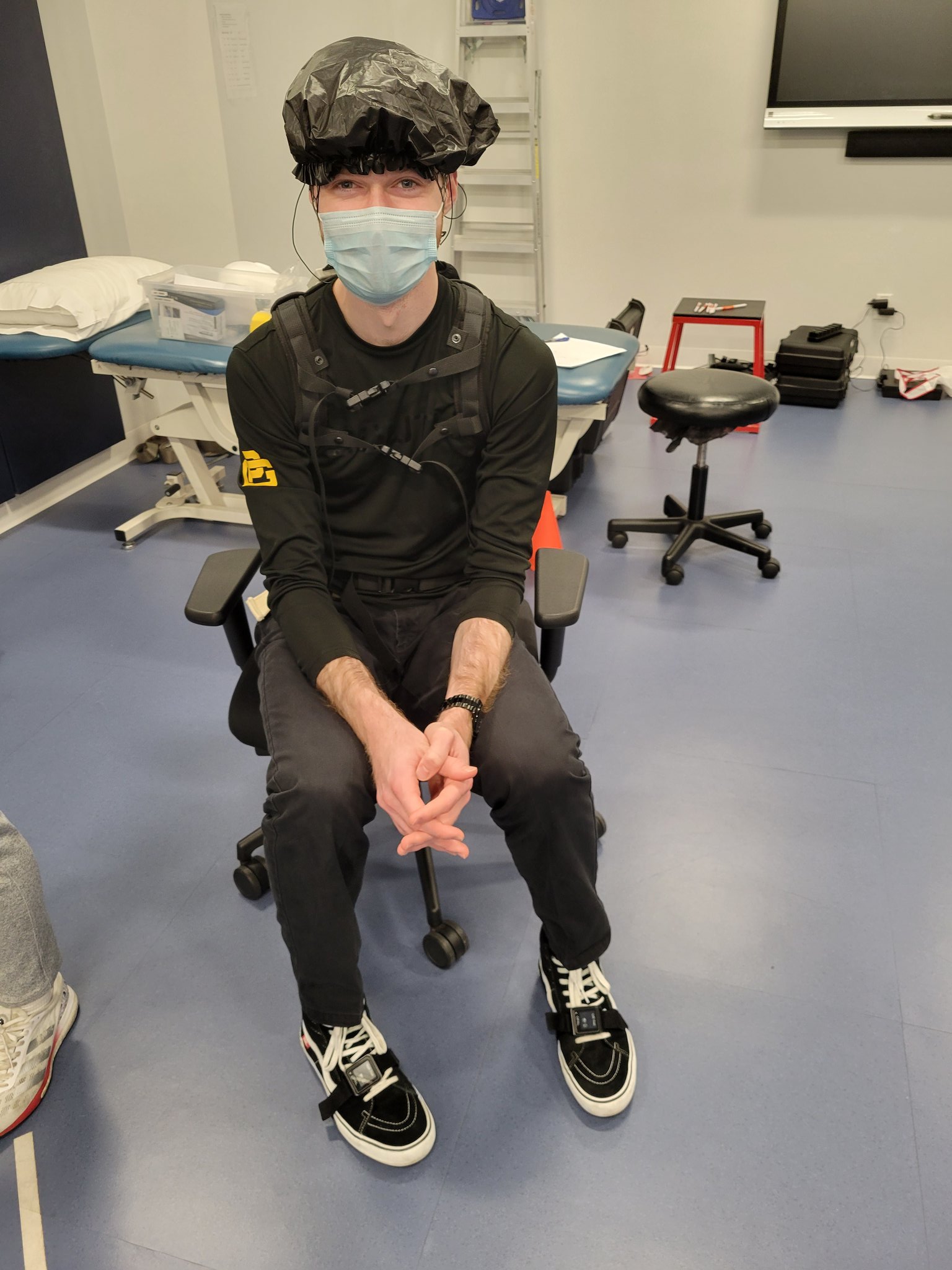
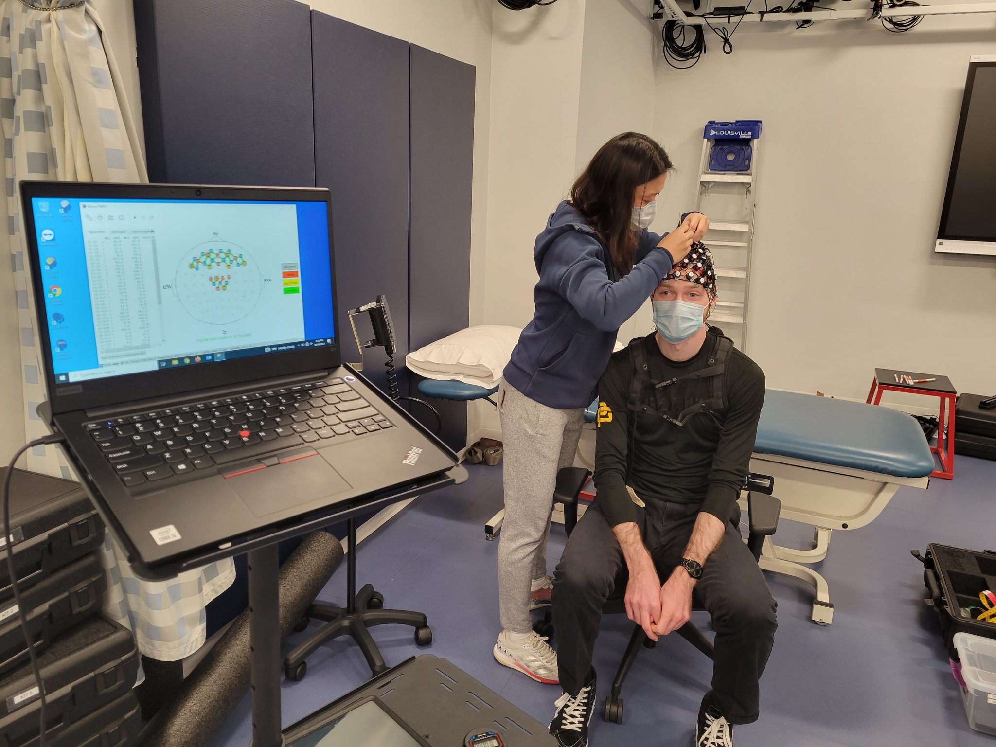
We use functional near-infrared spectroscopy (fNIRS) in collaboration with the Center of Neurophotonics for study of chronic pain and neuromotor control in people with knee osteoarthritis.


Musculoskeletal Imaging
MR imaging studies are performed at the new research-dedicated 3.0 Tesla MRI (Siemens PRISMA) at the Center for Integrated Life Sciences & Engineering (CILSE) located across the street from the Movement & Applied Imaging Lab. The scanner is equipped with an 18-channel surface coil for hip imaging and a 15-channel knee coil.
Radiography studies are performed at the outpatient Sports Medicine clinic at the Ryan Center, down the street from the Movement & Applied Imaging Laboratory. The BU Shuttle operates between the Ryan Center and the MAI Lab sites.
Cartilage MR Imaging

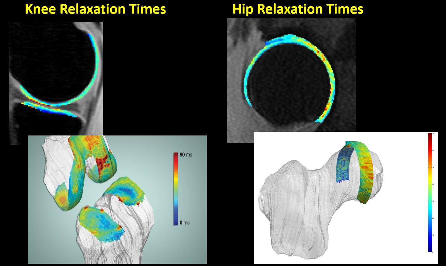
Advanced MR imaging is used to assess the morphology and composition of articular and meniscus cartilage. These include T1ρ and T2 relaxation times, and clinical scoring of cartilage and other articular tissues.
Muscle MR Imaging

Standard T1-w MR imaging is used to quantify muscle anatomical cross-sectional area. Water/fat MR imaging techniques are used to quantify the muscle adiposity.
Radiography
Clinical and research radiographic images are used to quantify hip and knee anatomy. Examples include Semi-flexed PA views for Kellgren-Lawrence grading, Long limb view for mechanical axis, and frog-leg views for hip alpha angle.
Strength and Physical Performance
Muscle Performance

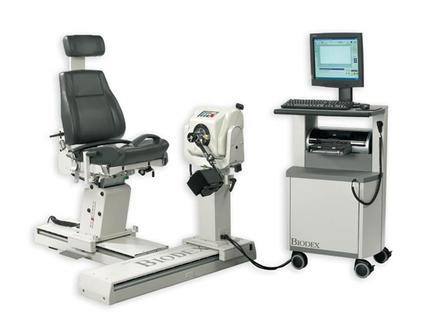
Muscle strength and endurance is assessed with a Biodex dynamometer (Biodex Medical Systems Inc.).
Physical Performance
Validated and population appropriate functional tests are used. Examples include single-leg hop for distance, timed up and go test, stair climbing test, 6 minute walk test, etc.
Digital Health Technologies
We are working with multiple technologies to capture physical activity, heart rate, sleep, and biomechanical data remotely from users in real-world environments.
We used REDCap and other software solutions to acquire patient-reported data at high temporal resolution for use in clinical trials for knee osteoarthritis.