Illustrated Dissector’s Manual
The opportunity to dissect in a cadaver lab is a profound educational experience that allows students to have insights into the human body in a way no textbook can provide. It builds both technical skills and anatomical knowledge while creating a deep appreciation for the donors who make this type of learning possible.
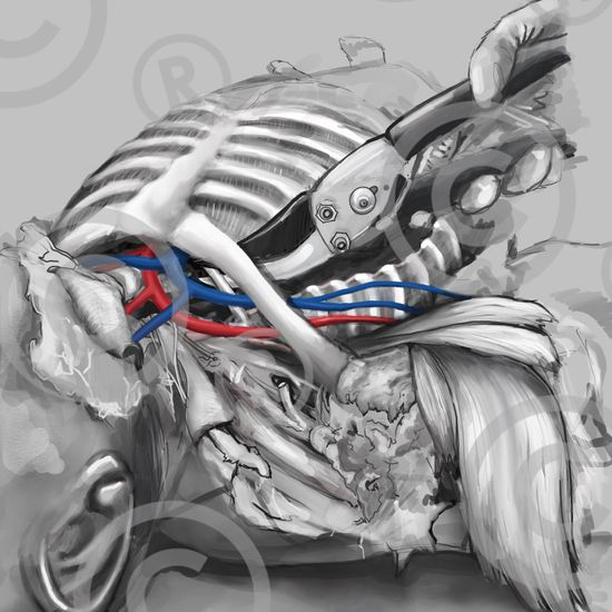
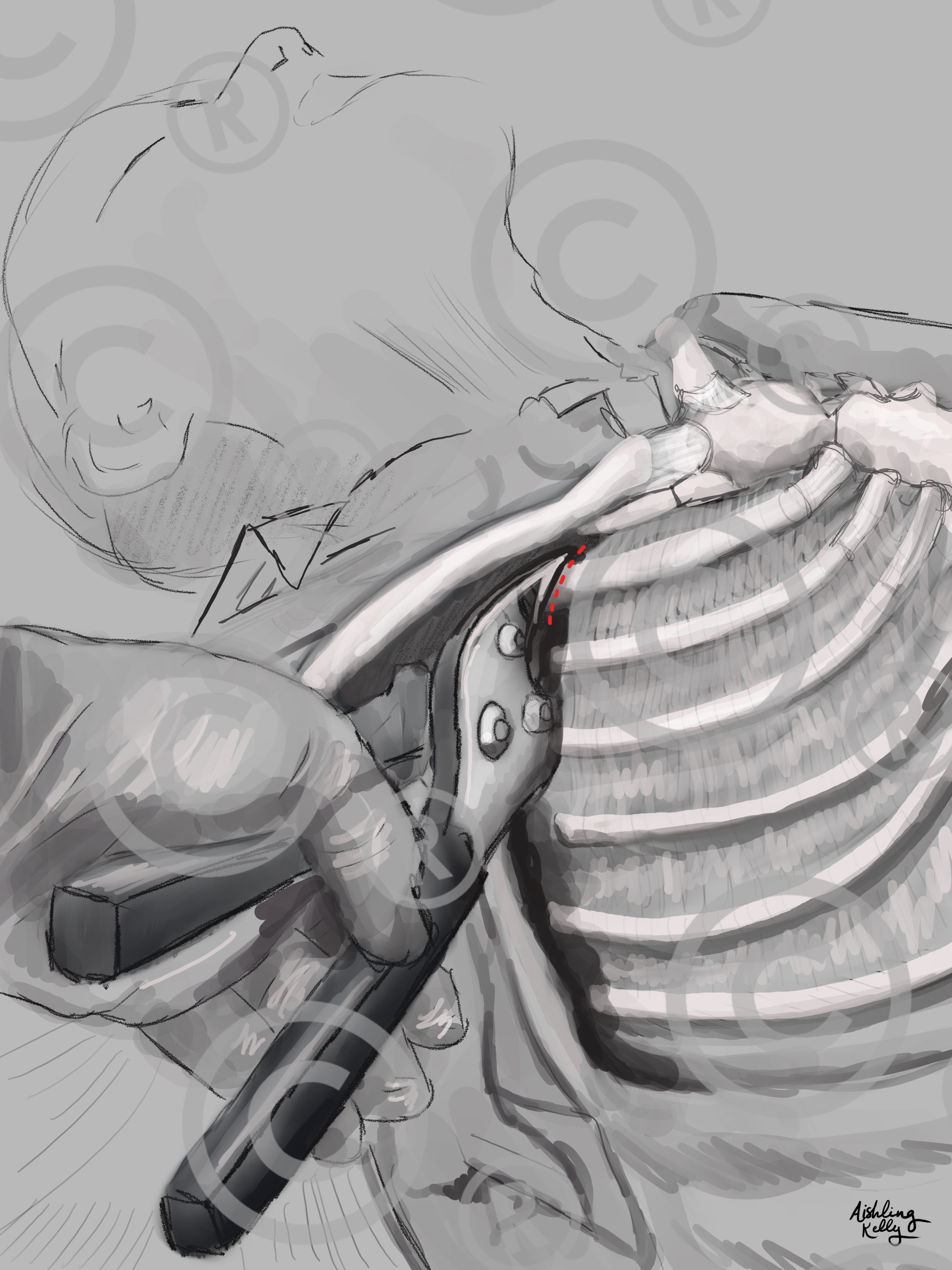
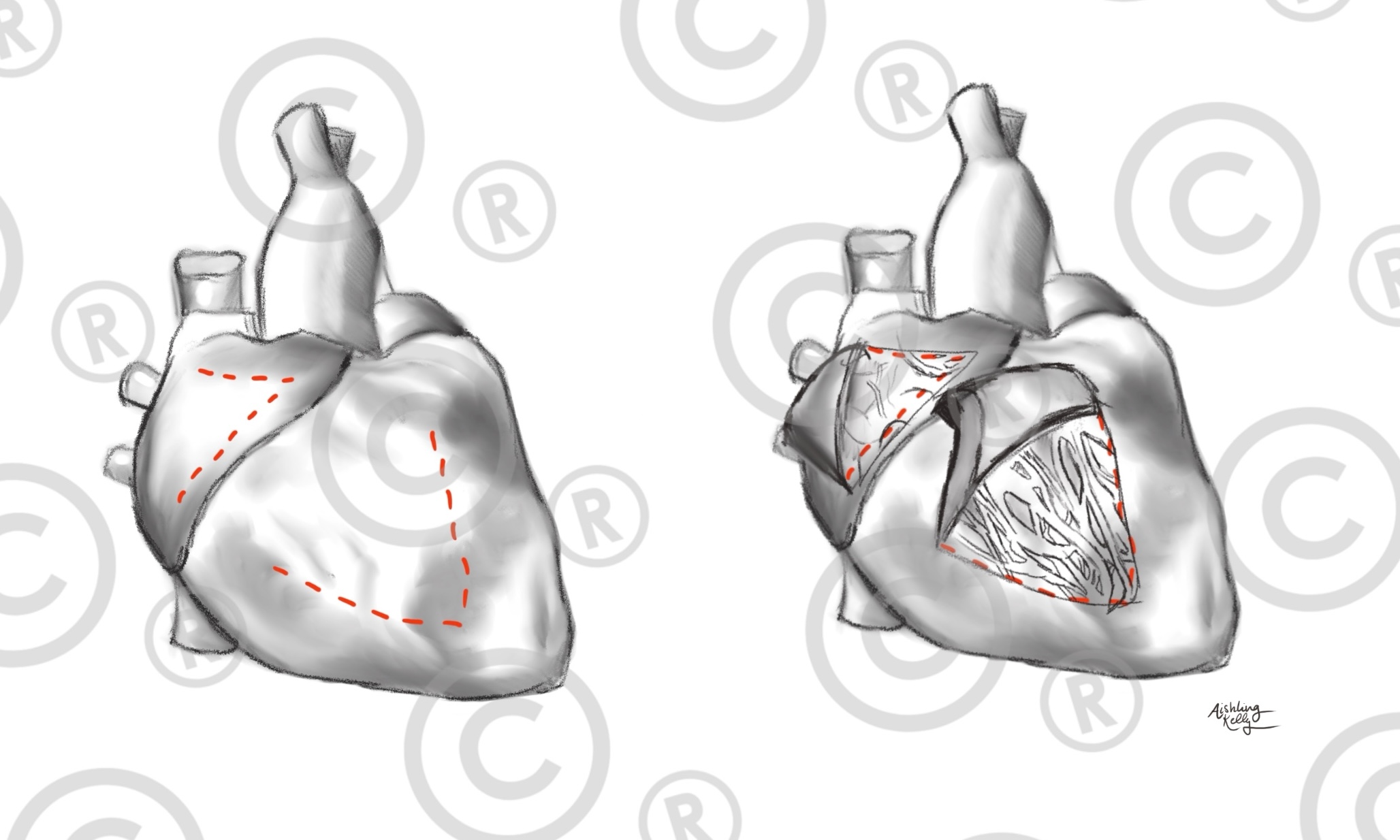
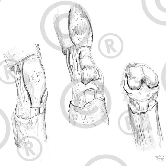
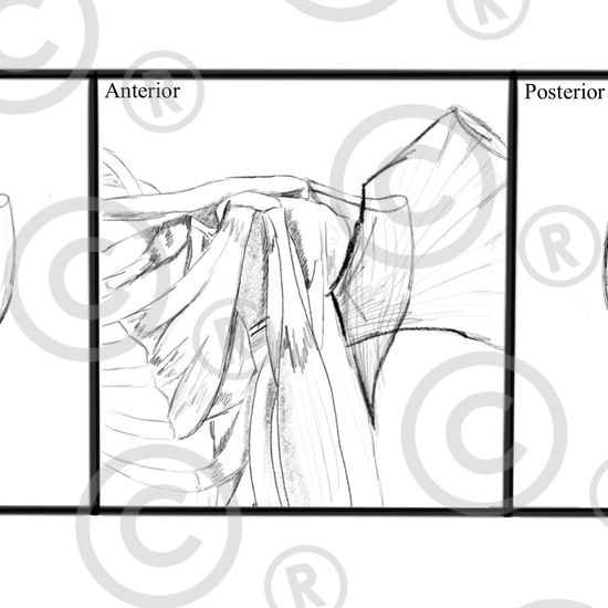
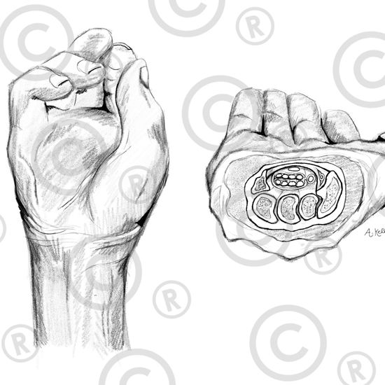
While textbooks and anatomy atlases provide a solid foundation, they often present idealized versions of the human body and rely heavily on dense text, making it challenging for dissectors to navigate structures effectively in the moment. The Illustrated Dissector’s Manual aims to bridge this gap by offering a comic strip-style guide that visually demonstrates the best cuts to highlight key anatomical features. Each illustration is drawn directly from donors and cross-referenced with Complete Anatomy to ensure accuracy. Our goal is to eventually publish a comprehensive Illustrated Dissector’s Manual to further support medical education.
If you are affiliated with Boston University (valid BU email address required), simply click on the infographic’s title link to be directed to Open BU where you can download the full-size image. If you are not affiliated with BU, please reach out to request access to full-size infographics.
This work is licensed under a Creative Commons Attribution-NonCommercial 4.0 International License. You are free to share and adapt the material for non-commercial purposes, with appropriate credit given.
For all inquiries, please visit our “Contact Us” page.
