News
Ribbon cutting ceremony celebrates the opening of the BU CryoEM Core Facility!
October 30, 2024

A ribbon cutting ceremony kicks off the official operations of the BU CryoEM Core Facility. Please reach out if you would like to get scheduled for an initial meeting.
BU CryoEM Core Facility workflow validation achieves a structure of apoferritin at 2.04 Å
August 16, 2024
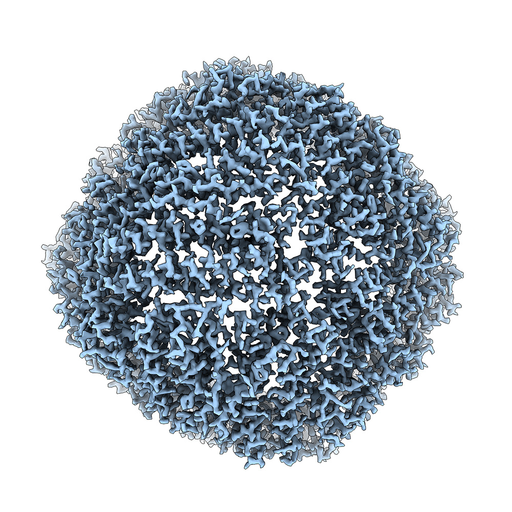
We validated our Glacios 2 microscope and Vitrobot Mark IV plunge-freezing sample preparation device for high-resolution data acquisition by freezing a sample of apoferritin and determining its structure to a resolution of 2.04 Å from a dataset of 930 exposures. Our external processing workstation running cryoSPARC LIVE provided a preliminary structure by processing data as it was being collected on the microscope. This fully integrated core facility test going from sample to structure validates the BU CryoEM Core Facility for user operation and high-quality data collection. Please reach out to us if you would like to receive training and reserve facility instrumentation.
The new cryo-EM Core is open for grid freezing and sample screening
July 22, 2024
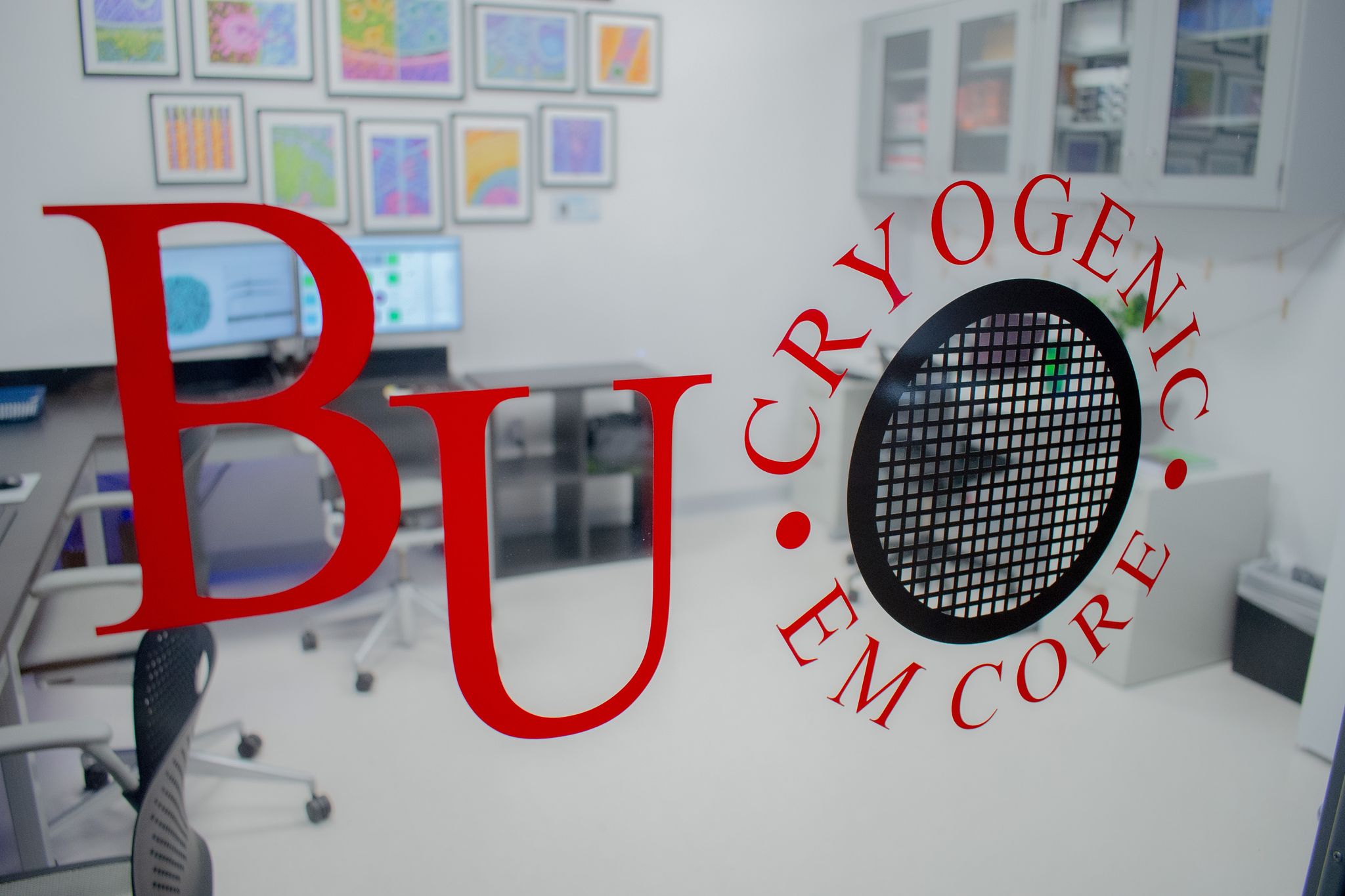
The new cryo-EM Core is open for grid freezing and sample screening! Fully automated data collection will be ready soon.
Celebrating the ‘First Light’ of the New CryoEM
April 19, 2024
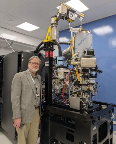
The ‘First Light’ of Boston University’s Glacios-2 cryogenic electron microscope (cryo-EM) is an exciting step in the setup of the new Cryo-EM Core Facility.
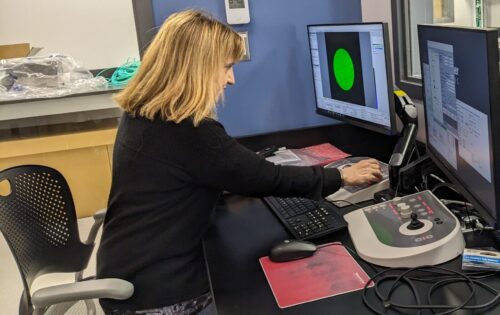
Located on the Medical Campus, there are 25 research groups across the university ready to use the new core, including teams from the dental and medical schools along with the College of Art & Sciences and College of Engineering.
With this new instrumentation, investigators will be able to advance scientific discoveries in health and disease by determining structures of macromolecules at near-atomic resolution. Samples are frozen so quickly that the water freezes as a glass, providing samples that are kept in a near-native state.
Thousands of images will be collected daily and analyzed using computers in the facility and at the Boston University Shared Computing Cluster. Planned experiments include studies on the heart, muscle, Alzheimer’s disease and other neurodegenerative disorders, G protein coupled receptor complexes, bacterial and viral infections, enzyme function, nanoparticles and protein biosynthesis and transport.
This is BU’s first cryo-EM core facility, updating older cryo-EMs located in the department of pharmacology, physiology & biophysics.
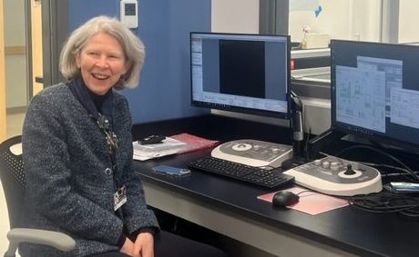
The cryo-EM was funded, in part, by a National Institutes of Health Shared Instrumentation Grant awarded to Professor of Pharmacology, Physiology & Biophysics Esther Bullitt, PhD. The expansion of EM facilities and recruitment of structural biology faculty has been one of the high-priority goals for Vanna Zachariou, PhD, Edward Avedisian Professor and chair of the department.
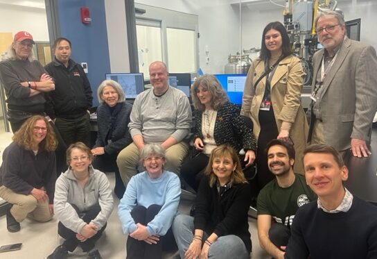
A ‘First Light’ gathering was held Friday to celebrate the inaugural electron beam from the instrument, which was configured by Dean Antman. Alignment and testing of the Glacios-2 are continuing, and it is expected the facility will be ready for use in the near future.
https://www.bumc.bu.edu/camed/2024/04/19/celebrating-the-first-light-of-the-new-cryoem/
Glacios-2 cryo-TEM has landed at Boston University
March 15, 2024
We have received the shipment of crates containing the Glacios-2 cryo transmission electron microscope and supporting equipment. The components will be carefully assembled and aligned over the next few months by ThermoFisher to prepare for high quality data collection in a semi-automated high-throughput fashion. We are very excited for all of the future discoveries to come out of this cryoEM core facility!