Partitioned aperture wavefront imaging
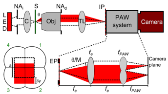 A wavefront imager provides images of the phase and amplitude of a wavefront. Several techniques for wavefront imaging have been developed. Most of these involve the use of a laser, and are thus susceptible to speckle noise. Techniques that do not require a laser generally require collimated light, meaning they are not light efficient, or require moving parts and the acquisition of multiple images, meaning they are slow. We have recently developed a wavefront imager that is fast (single shot), achromatic (works with broadband light), light efficient (works with extended sources), and simple. The technique is based on partitioning the detection aperture of a standard microscope into four quadrants with the use of four off-axis lens. These lenses provide four oblique detection images that are simultaneously acquired with a single camera. The data provided by these four images enables the reconstruction of wavefront phase and amplitude with a simple numerical algorithm that runs in real time (video rate).
- R. Barankov, J.-C. Baritaux, J. Mertz, “High-resolution 3D phase imaging using a partitioned detection aperture: a wave-optic analysis”, J. Opt. Soc. Am. A. 32, 2123-2135 (2015). link
- J.-C. Baritaux, C. R. Chan, J. Li, J. Mertz, “View synthesis with a partitioned-aperture microscope”, Opt. Lett. 39, 685-88 (2014). link
- R. Barankov and J. Mertz, “Single-exposure profilometry using partitioned aperture wavefront imaging”, Opt. Lett. 38, 3961-3963 (2013). link
- B. Parthasarathy, K. K. Chu, T. N. Ford, J. Mertz, “Quantitative phase imaging using a partitioned detection aperture”, Opt. Lett. 37, 4062-4064 (2012). link
- K. K. Chu, J. Mertz, “Single exposure complementary aperture phase microscopy with polarization encoding”, Opt. Lett. 37, 3798-3800 (2012). link