Nonlinear microscopy

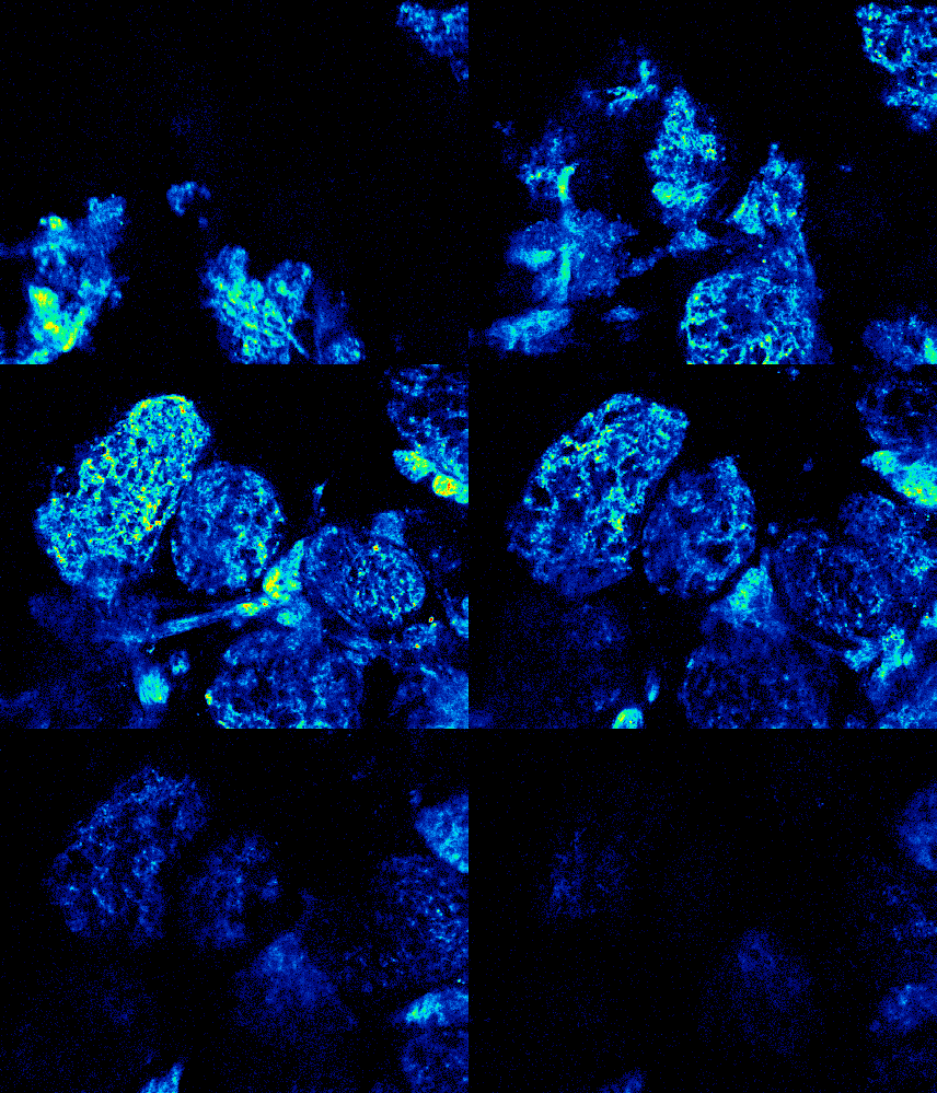
Nonlinear optical microscopy is generally divided into two categories: incoherent or coherent. Incoherent microscopy produces an optical signal whose phase is random and whose power is proportional to the concentration of radiating molecules. Fluorescence is a common example of an incoherent signal. Nonlinear versions of fluorescence microscopes are based on the simultaneous absorption of two or more photons, the most well known being two-photon excited fluorescence (TPEF) microscopy. In TPEF microscopy, two excitation photons from a pulsed laser (typically a Ti:sapphire laser of wavelength 700nm-1000nm) combine to excite a fluorescent molecule. The molecule then releases its excitation energy as a fluorescence photon (typically a visible wavelength). Because the excitation is nonlinear, the fluorescence is confined to the focal center of the laser beam, and fluorescence power decays as 1/z2, where z is the axial distance away from the focus (see figure). TPEF microscopy therefore confers 3D-imaging with out-of-focus background rejection similar to a confocal microscope. The advantage of TPEF microscopy over confocal microscopy is that it can penetrate deeper in thick tissue.
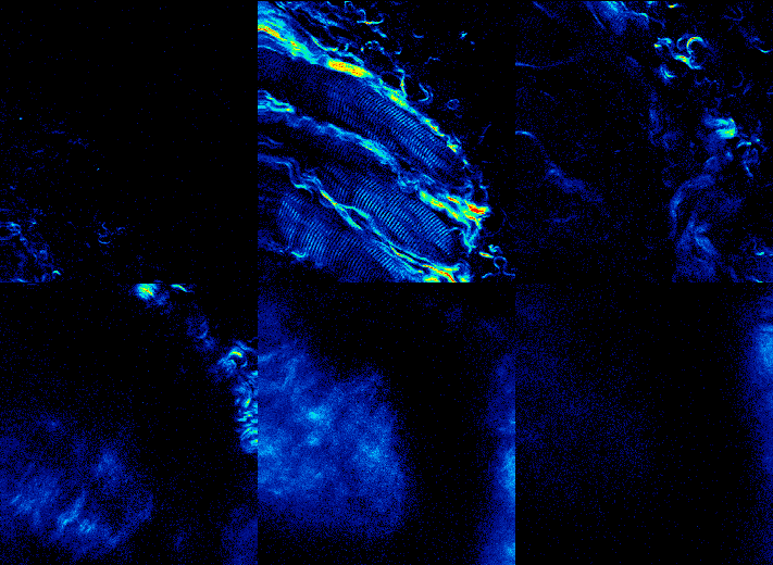
Coherent microscopes produce optical signals whose phase is rigorously prescribed by a variety of factors including the excitation light phase and the geometric distribution of the radiating molecules. Coherent signal power is proportional to the concentration of radiating molecules squared. Nonlinear versions of coherent microscopy are based on the simultaneous scattering of two or more photons. Examples are second-harmonic generation (SHG) and coherent anti-Stokes Raman scattering (CARS) microscopy. SHG signals are highly sensitive to molecular orientation.
We have worked on further developments of TPEF microscopy and its applications to brain imaging. For example, we have implemented TPEF microscopy with simultaneous autoconfocal microscopy, differential aberration imaging (DAI), and reverberation multiplane imaging. We have also implemented an optical dynamic clamp for calcium uncaging.

- E. Idoux and J. Mertz, “Control of local intracellular calcium concentration with dynamic-clamp controlled 2-photon uncaging”, PLoS ONE, 6, e28685 (2011). link
- K. Lillis, A. Eng, J. White, J. Mertz, “Two-photon imaging of spatially extended neuronal network dynamics with high temporal resolution”, J. Neurosci. Meth. 172, 178-184 (2008). link
- A. Leray, K. Lillis, J. Mertz, “Enhanced background rejection in thick tissue with differential-aberration two-photon microscopy”, Biophys. J. 94, 1449-1458 (2008). link
- K. K. Chu, D. Lim, J. Mertz, “Enhanced weak-signal sensitivity in two-photon microscopy by adaptive illumination”, Opt. Lett. 32, 2846-2848 (2007). link
- A. Leray, J. Mertz, “Rejection of two-photon fluorescence background in thick tissue by differential aberration imaging”, Opt. Express 14, 10565-10573 (2006). link
- T. Pons, J. Mertz, “Membrane potential detection with second-harmonic generation and two-photon excited fluorescence: a theoretical comparison”, Opt. Commun. 258, 203-209 (2006). link
- J. Mertz, “Nonlinear microscopy: new techniques and applications”, Curr. Opin. Neuro. 14, 610-616 (2004). link
- L. Porrès, O. Mongin, C. Katan, M. Charlot, T. Pons, J. Mertz, M. Blanchard-Desce, “Enhanced two-photon absorption with novel octupolar propeller-shaped fluorophores derived from triphenylamine”, Org. Lett. 6, 47-50 (2004). link
- T. Pons, L. Moreaux, O. Mongin, M. Blanchard-Desce, J. Mertz, “Mechanisms of membrane potential sensing with second harmonic generation microscopy”, J. Biomed. Opt. 8, 428-431 (2003). link
- L. Moreaux, T. Pons, M. Blanchard-Desce, J. Mertz, “Electro-optic response of second-harmonic generation membrane potential sensors”, Opt. Lett. 25, 625-627 (2003). link