Graded field microscopy
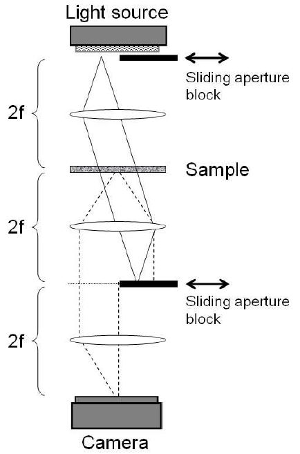
Graded-field microscopy is a general technique for obtaining phase-gradient contrast in biological tissue slices. The technique is based on introducing partial beam blocks in the illumination and detection apertures of a standard white-light widefield transillumination microscope. Depending on the relative aperture sizes, one block produces phase-gradient contrast while the other reduces brightfield background, allowing a full operating range between brightfield and darkfield contrast.