Active-illumination microscopy
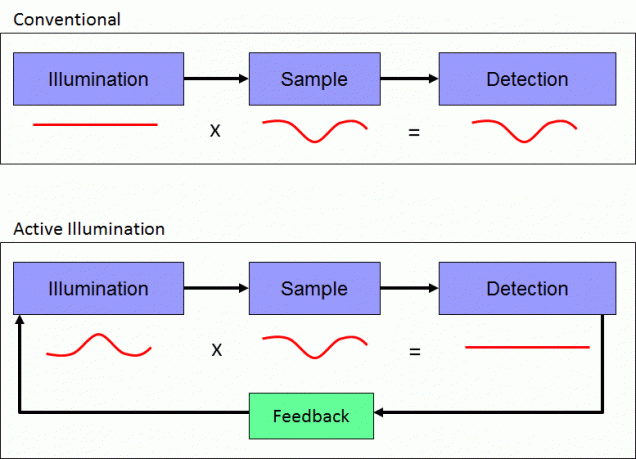
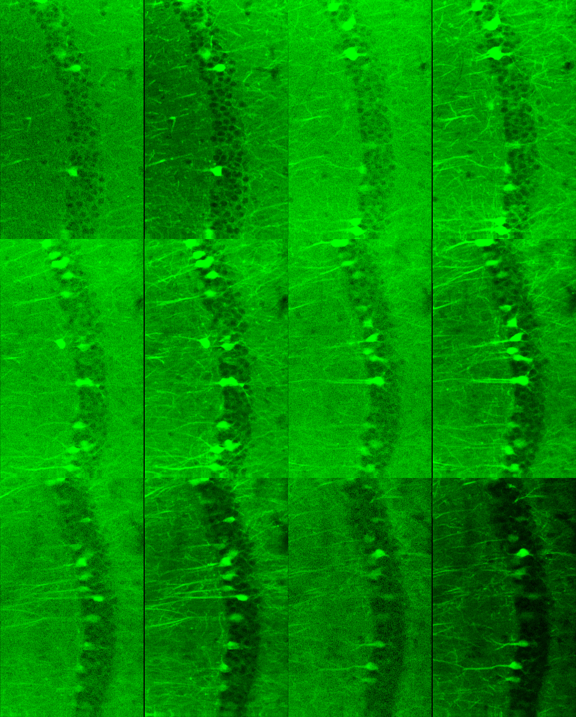
 Active illumination microscopy (AIM) is a method of redistributing dynamic range in a scanning microscope using real-time feedback to control illumination power on a sub-pixel time scale. The principle of the feedback is to maintain a fixed detection power, thereby transferring high measurement resolution to weak signals while virtually eliminating the possibility of image saturation. As a result, this technique enhances both the weak-signal relative sensitivity and the dynamic range of a laser scanning optical microscope. We have demonstrated a fully integrated instrument that performs both feedback and image reconstruction. The image is reconstructed on a logarithmic scale to accommodate the dynamic range benefits of AIM in a single output channel. While AIM is applicable to any type of scanning microscope, we have applied it specifically to two-photon microscopy.

- R. Yang, T. D. Weber, E. D. Witkowski, I. G. Davison, J. Mertz, “Neuronal imaging with ultrahigh dynamic range multiphoton microscope”, Sci. Rep. 7:5817 (2017). link
- K. K. Chu, D. Lim, J. Mertz, “Practical implementation of log-scale active illumination microscopy”, Biomed. Opt. Express, 1, 236-245 (2010). link
- K. K. Chu, D. Lim, J. Mertz, “Enhanced weak-signal sensitivity in two-photon microscopy by adaptive illumination”, Opt. Lett. 32, 2846-2848 (2007). link